TRAUMA UROGENITAL
890 likes | 1.04k Vues
TRAUMA UROGENITAL. Anatomi Ginjal Ureter Buli buli Uretra. TRAUMA UROGENITAL. Anatomi Ginjal Ureter Buli buli Uretra. Ginjal. Sepasang organ seperti kacang Terletak retroperitonel diregio lumbal superior Dilapisi oleh 3 lapis jaringan penunjang: kapsul ginjal

TRAUMA UROGENITAL
E N D
Presentation Transcript
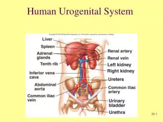
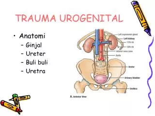
TRAUMA UROGENITAL • Anatomi • Ginjal • Ureter • Buli buli • Uretra
TRAUMA UROGENITAL • Anatomi • Ginjal • Ureter • Buli buli • Uretra
Ginjal • Sepasang organ seperti kacang • Terletak retroperitonel diregio lumbal superior • Dilapisi oleh 3 lapis jaringan penunjang: • kapsul ginjal • kapsul adipose dan • fasia renalis
Ureter • Tubulus muscular yang menghubungkan ginjal ke buli buli • Terletak di belakang rongga peritoneum (retroperitoneal) • Panjang 25 – 30 cm
TRAUMA GINJAL Trauma Ginjal • Sering • 8-10% trauma tumpul / tajam abdomen • Separuh dari kejadian trauma urogenital • Di proteksi : * Otot-otot lumbal * Iga * Vertebrae
Angka kesakitan / kematian ok trauma ginjal tergantung : • Derajat trauma • Keterlibatan trauma organ lain • Fasilitas penanggulangan trauma
Buli-buli • Buli buli normal dapat menampung 350 – 450 mL urine • Drainase kendung kemih bermuara ke vena iliaca interna
Uretra • Tabung yang menyalurkan urine ke luar dari buli-buli • Secara anatomis uretra dibagi menjadi 2 bagian : • Uretra posterior dan • Uretra anterior
Mekanisme Trauma Trauma tumpul -> penyebab trauma Langsung, tidak langsung Trauma tumpul langsung • KLL • Olah raga • Kecelakaan kerja • Perkelahian
Trauma tumpul tidak langsung * Jatuh dari ketinggian * KLL menyebabkan pergerakan ginjal tiba-tiba dlm rongga retro peritonium Avulsi pedikel ginjal Robekan tunika intima
Bisa juga oleh trauma iatrogenik • Pemasangan kateter di atas ureter • Pengambilan biopsi ginjal • Infeksi tidak langsung Klasifikasi * Ada beberapa macam * Ditentukan oleh luas dan penatalaksanaan
Cedera Ginjal * Minor * Mayor * Vaskuler Cedera Minor • 90% trauma ginjal • Kontusio ginjal • Laserasi parenkim superficial
Cedera Mayor • Laserasi korteks, medula tanpa ekstravasasi • Laserasi korteks, medula dengan ekstravasasi Cedera Vaskuler • Avulsi • Trombosis
Berdasarkan AAST( American for The Surgery of Trauma ) Dibagi 5 derajat Derajat 1 • Kontusio ginjal /subkapsularhematom • Tidak meluas • Hematuria dengan normal imaging
Derajat 2 • Hematom perineal • Tdk meluas ke retroperitonium • Laserasi superficial ( < 1cm ) • Tdk melibatkan collecting systim Derajat 3 • Renal laserasi ( > 2cm ) • Sub capsular hematom • Perinephric hematom • Tdk melibatkan collecting systim
Derajat 4 • Laserasi yang meluas ke collecting systim • Extravasasi • Trauma vasculer segmental infark
Derajat 5 • Shattered kidney • Devaskularisasi / oklusi / trombosis arteri / vena utama • Laserasi komplit • Extravasasi • UPJ avulsi
Pemeriksaan Radiologi • Foto polos abdomen • IVP ( Intra Vena Pyelografi ) • USG ( Ultra Sonographi ) • CT Scan abdomen / Whole abdomen • uretrocistografi
IVP* Melihat ekstravasasi urin / kontras* Tidak bisa mendeteksi trauma ginjal derajat I, II* Fungsi ginjal kontra lateralUSG* Melihat hemoperitoneum* Tdk dianjurkan utk evaluasi trauma ginjal* Dengan color doppler melihat vaskuler
CT Scan • Pemeriksaan yang sensitif dan spesifik • Menentukan derajat trauma • Tidak invasif • Dpt mengevaluasi organ lain ( hepar , lien , aorta ) kontras non kontras Angiografi • Invasif • Delayed renal bleeding-pseudo-aneurisma
Gambar 1. Kidney trauma. Absent nephrogram. Abdominal radiograph after intravenous contrast administration in a patient with hypotension after a motor vehicle collision shows absent right nephrogram
Gambar 2. Kidney trauma. Grade 3 renal laceration on abdominal radiograph. Abdominal radiograph after intravenous contrast administration shows very diminished left nephrogram and no urinary contrast extravasation
Gambar 3. Kidney trauma. Grade 5 renal injury. Shattered kidney with renal vein thrombosis (incomplete). Abdominal radiograph after intravenous contrast administration shows absent right nephrogram
Gambar 4 Kidney trauma. Grade 1 renal injury, contusion. Image from a contrast-enhanced CT scan of the abdomen in a patient with hematuria after a motor vehicle collision shows ill-defined area of hypoenhancement in the medial right kidney.
Gambar. 5. Kidney trauma. Grade 1 renal injury, subcapsular hematoma. CT scan of the abdomen with intravenous contrast in a patient after a motor vehicle collision shows crescentic high-density fluid collection around the left kidney. Note the well-defined outer margin
Gambar 6. Kidney trauma. Grade 1 renal injury, subcapsular hematoma. CT scan of the abdomen with intravenous contrast in a patient after a motor vehicle collision; shows crescentic high-density fluid collection around the left kidney. Note the well-defined outer margin and the mild deformity of the renal parenchyma
Derajat II dan III Gambar 6. Kidney trauma. Grade 2 renal injury, subcapsular and perinephric hematomas. Contrast-enhanced CT scan of the abdomen on a patient with hematuria after a motor vehicle collision shows an ill-defined fluid collection in the left perinephric space. There is also a subcapsular hematoma with deformity of the renal parenchyma
Gambar 7 Kidney trauma. Grade 2 renal injury, perinephric hematoma. Contrast-enhanced CT scan of the abdomen on a patient with hematuria after a motor vehicle collision shows an ill-defined fluid collection in the left perinephric space
Gambar 8. Kidney trauma. Grade 3 renal laceration with normal one-shot intravenous pyelogram. CT scan through the kidneys after intravenous contrast on the same patient as in Image 1 shows renal laceration and perinephric hematoma.
Gambar 9 Kidney trauma. Grade 2 renal laceration. Contrast-enhanced CT scan of the abdomen after a motor vehicle collision shows a superficial (less than 1 cm deep) renal parenchymal defect with a large perinephric hematoma
Gambar 10. Kidney trauma. Grade 2 renal laceration. Delayed image shows no urinary contrast extravasation. Contrast-enhanced CT scan of the abdomen after a motor vehicle collision shows a superficial (<1 cm deep) renal parenchymal defect with a large perinephric hematoma
Gambar 11. Kidney trauma. Grade 3 renal laceration. CT scan of the abdomen after intravenous contrast administration shows irregular nonenhancing renal parenchymal defect with extension greater than 1 cm deep to near the renal pelvis. no urinary contrast extravasation
Gambar 12. Kidney trauma. Grade 3 renal laceration. CT scan of the abdomen after intravenous contrast administration shows irregular nonenhancing renal parenchymal defect with extension greater than 1 cm deep to near the renal pelvis. This delayed image showed no urinary contrast extravasation.
Derajat IV Gambar13 Kidney trauma. Grade 4-5 renal injury. Lacerations extending to the collecting system. Contrast-enhanced CT scan of the abdomen in a patient with hematuria after a motor vehicle collision shows deep lacerations extending into the collecting system of the right kidney. Extension into the collecting system is confirmed by urinary contrast extravasation on delayed image through the kidney in excretory phase
Gamba14. Kidney trauma. Grade 4-5 renal injury. Lacerations extending to the collecting system. Contrast-enhanced CT scan of the abdomen in a patient with hematuria after a motor vehicle collision shows deep lacerations extending into the collecting system of the right kidney (Image 22). Extension into the collecting system is confirmed by urinary contrast extravasation on this delayed image through the kidney in excretory phase
Gambar 15. Kidney trauma. Grade 4 renal injury segmental infarction. Contrast-enhanced CT scan of the upper abdomen shows a segmental area of nonenhancement in the upper medial left kidney without associated renal laceration
Gambar 16. Kidney trauma. Grade 4 renal injury segmental infarction. Contrast-enhanced CT scan of the upper abdomen in another patient after a motor vehicle collision shows a segmental area of nonenhancement in the upper medial left kidney without associated renal laceration
Derajat V Gambar 17. Kidney trauma. Grade 5 renal injury. Shattered kidney. Contrast-enhanced CT scan of the abdomen in a patient with hematuria and hypotension after a motor vehicle collision shows transection of the right kidney with a large hematoma around and between the 2 halves of the kidney. The 2 halves are both perfused because there were 2 renal arteries Delayed images show urinary contrast extravasation
Gambar 18. Kidney trauma. Grade 5 renal injury. Shattered kidney. Contrast-enhanced CT scan of the abdomen in a patient with hematuria and hypotension after a motor vehicle collision shows transection of the right kidney with a large hematoma around and between the 2 halves of the kidney. The 2 halves are both perfused because there were 2 renal arteries. Delayed images show urinary contrast extravasation
Gambar 19 Kidney trauma. Grade 5 renal injury. Shattered kidney. Contrast-enhanced CT scan of the abdomen in a patient with hematuria and hypotension after a motor vehicle collision shows transection of the right kidney with a large hematoma around and between the 2 halves of the kidney. The 2 halves are both perfused because there were 2 renal arteries Delayed images show urinary contrast extravasation
Gambar 20. Kidney trauma. Grade 5 renal injury. Shattered kidney. Contrast-enhanced CT scan of the abdomen in a patient with hematuria and hypotension after a motor vehicle collision shows transection of the right kidney with a large hematoma around and between the 2 halves of the kidney. The 2 halves are both perfused because there were 2 renal arteries. Delayed images show urinary contrast extravasation
Gambar 21. Kidney trauma. Grade 5 renal injury. Shattered kidney with renal vein thrombosis (incomplete). CT scan of the abdomen with intravenous contrast administration shattered right kidney and renal vein thrombus extending slightly into the inferior vena cava
Gambar 22. Kidney trauma. Normal ultrasound with grade 5 renal injury. Ultrasound gray-scale image of a patient involved in a motor vehicle collision shows what appears to be a normal right kidney
Gambar 23 Kidney trauma. Grade 5 renal injury. Color Doppler ultrasound of same motor vehicle collision patient as in Image 4 shows no blood flow within the right kidney.
4.Arteriografy Gambar 24. Kidney trauma. Active vascular contrast extravasation. Catheter angiography during arterial phase on the same patient as in Image 40 shows a small pseudoaneurysm at the lower pole
gambar 25. Kidney trauma. Active vascular contrast extravasation. Catheter angiography during nephrographic phase in the same patient as in Image 41 shows a small pseudoaneurysm at the lower pole