Membrane Transport
600 likes | 653 Vues
Learn how solutes cross cell membranes, factors affecting movement, comparison of transport mechanisms, ion channels function, and epithelial transport processes. Explore importance in physiological functions, diseases, and therapeutic strategies.

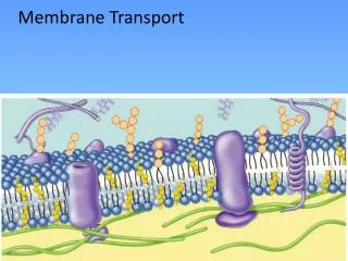
Membrane Transport
E N D
Presentation Transcript
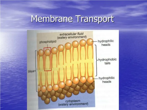
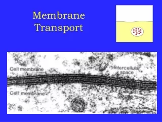
Membrane Transport Xia Qiang(夏强), PhD Department of Physiology Zhejiang University School of Medicine Tel: 88206861 Email: xiaqiang@zju.edu.cn
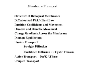
LEARNING OBJECTIVES • Describe how solutes cross cell membranes • Explain how charge, size, and solubility affect solute movement across cell membranes • Contrast how transporters, pumps & channels work • Describe how ion channels are gated • Describe how epithelial transport works
Membrane Transport • Simple Diffusion(单纯扩散) • Facilitated Diffusion(易化扩散) • Active Transport(主动转运) • Endocytosis and Exocytosis(出胞与入胞)
Importance of pumps, transporters & channels • Basis of physiologic processes • Growth • Metabolic activities • Sensory perception • Basis of disease • Defective transporters (cystic fibrosis) • Defective channels (long QT syndrome, paralysis) • Basis of pharmacological therapies • Hypertension (diuretics) • Stomach ulcers (proton pump inhibitors)
START: Initially higher concentration of molecules randomly move toward lower concentration. Over time, solute molecules placed in a solvent will evenly distribute themselves. Diffusional equilibrium is the result (Part b).
At time B, some glucose has crossed into side 2 as some cross into side 1.
Note: the partition between the two compartments is a membrane that allows this solute to move through it. Net flux accounts for solute movements in both directions.
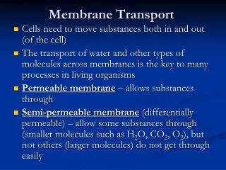
Simple Diffusion Relative to the concentration gradient movement is DOWN the concentration gradient ONLY (higher concentration to lower concentration) Rate of diffusion depends on • The concentration gradient • Charge on the molecule • Size • Lipid solubility
Facilitated Diffusion • Carrier-mediated
Characteristics of carrier-mediated diffusion net movement always depends on the concentration gradient • Specificity • Saturation • Competition
Channel-mediated 3 cartoon models of integral membrane proteins that function as ion channels; the regulated opening and closing of these channels is the basis of how neurons function.
The opening and closing of ion channels results from conformational changes in integral proteins. Discovering the factors that cause these changes is key to understanding excitable cells.
Regulation of gating in ion channels. Several types of gating are shown for ion channels. A) Ligand-gated channels open in response to ligand binding. B) Protein phosphorylation or dephosphorylation regulate opening and closing of some ion channels. C) Changes in membrane potential alter channel openings. D) Mechanical stretch of the membrane results in channel opening.
Different ways in which ion channels form pores. Many K+ channels are tetramers A), with each protein subunit forming part of the channel. In ligand-gated cation and anion channels B) such as the acetylcholine receptor, five identical or very similar subunits form the channel. Cl- channels from the ClC family are dimers C), with an intracellular pore in each subunit. Aquaporin water channels (D) are tetramers with an intracellular channel in each subunit.
Diagrammatic representation of the pore-forming subunits of three ion channels. The α subunit of the Na+ and Ca2+ channels traverse the membrane 24 times in four repeats of six membrane-spanning units. Each repeat has a “P” loop between membrane spans 5 and 6 that does not traverse the membrane. These P loops are thought to form the pore. Note that span 4 of each repeat is colored in red, representing its net “+” charge. The K+ channel has only a single repeat of the six spanning regions and P loop. Four K+ subunits are assembled for a functional K+ channel.
Three types of passive, non-coupled transport through integral membrane proteins
membrane In both simple and facilitated diffusion, solutes move in the direction predicted by the concentration gradient. In active transport, solutes move opposite to the direction predicted by the concentration gradient.
Active transport • Primary active transport(原发性主动转运) • Secondary active transport(继发性主动转运)
Primary Active Transport making direct use of energy derived from ATP to transport the ions across the cell membrane
Here, in the operation of the Na+-K+-ATPase, also known as the “sodium pump,” each ATP hydrolysis moves three sodium ions out of, and two potassium ions into, the cell.
The ion gradients established by primary active transport permits the transport of other substances against their concentration gradients Secondary Active Transport
Cotransport(同向转运) the ion and the second solute cross the membrane in the same direction (e.g. Na+-glucose, Na+-amino acid cotransport) Countertransport(逆向转运) the ion and the second solute move in opposite directions (e.g. Na+-Ca2+, Na+-H+ exchange)
Composite diagram of main secondary effects of active transport of Na+ and K+
Osmosis(渗透) Solvent + Solute = Solution Here, water is the solvent. The addition of solute lowers the water concentration. Addition of more solute would increase the solute concentration and further reduce the water concentration.
Begin: The partition between the compartments is permeable to water and to the solute. After diffusional equilibrium has occurred: Movement of water and solutes has equalized solute and water concentrations on both sides of the partition.
Begin: The partition between the compartments is permeable to water only. After diffusional equilibrium has occurred: Movement of water only has equalized solute concentration.
Alternative functions of endocytosis: • Transcellular transport 2. Endosomal processing 3. Recycling the membrane 4. Destroying engulfed materials
Endocytosis • Phagocytosis • Pinocytosis • Clathrin-mediated endocytosis • Caveolae-dependent uptake • Nonclathrin/noncaveolae endocytosis
Two pathways of exocytosis • Constitutive exocytosis pathway -- Many soluble proteins are continually secreted from the cell by the constitutive secretory pathway • Regulated exocytosis pathway -- Selected proteins in the trans Golgi network are diverted into secretory vesicles, where the proteins are concentrated and stored until an extracellular signal stimulates their secretion
Steps to exocytosis • Vesicle trafficking: In this first step, the vesicle containing the waste product or chemical transmitter is transported through the cytoplasm towards the part of the cell from which it will be eliminated • Vesicle tethering: As the vesicle approaches the cell membrane, it is secured and pulled towards the part of the cell from which it will be eliminated • Vesicle docking: In this step, the vesicle comes in contact with the cell membrane, where it begins to chemical and physically merge with the proteins in the cell membrane • Vesicle priming: In those cells where chemical transmitters are being released, this step involves the chemical preparations for the last step of exocytosis • Vesicle fusion: In this last step, the proteins forming the walls of the vesicle merge with the cell membrane and breach, pushing the vesicle contents (waste products or chemical transmitters) out of the cell. This step is the primary mechanism for the increase in size of the cell's plasma membrane