Download
1 / 26
260 likes | 285 Vues
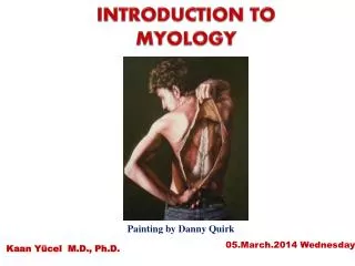
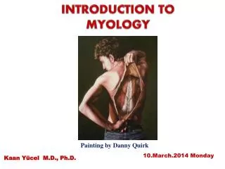
Explore the intricate world of myology, including muscle functions, types, structures, and divisions. Learn about the role muscles play in locomotion, thermoregulation, and communication.

E N D
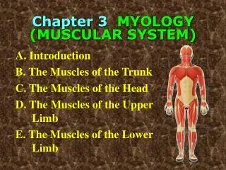
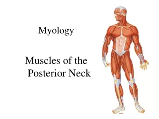
GENERAL MYOLOGY (Muscles - an active part of the locomotor system)
General function of muscles *produce movement in sites of skeletal junctions * change shapes of various body cavities and openings * give information about the body position in 3D space * important role during thermoregulation * help toblood and lymphcirculation * verbal and non verbal comunication * about 600 muscles (♂ 35%, ♀ 32% of weigth) * logistic system (supports respiration, digestion…) Smooth musculature forms walls of vessels and hollow organs, works without our will, without fatigue Srdeční svalovina tvoří srdce, pracuje bez závislosti na naší vůli, po celý život, bez únavy Svalovina příčně pruhovaná, tvoří kosterní svalstvo, pracuje v závislosti na naší vůli, snadno se unaví, spotřebuje hodně energie, při práci tvoří teplo
On the basis of structure and physiologicalcharacteristics we distinguish: • Striated (skeleton) muscles– musculi sceleti (form muscles of limbs, work under control of our will, easy fatigued, spend a lot of energy, produce heat) + skin muscles(musculi cutanei) 2) Cardiac muscles (myocardium) 3) Non-striatedvisceral (smooth) muscles– form an integralpart of some hollow organs and cavities - work without ourwill, without fatigue).
The main of the mechanical function of muscle fibers is shortening - contraction (movement).
Common structure of muscle Origo (origin) Fascia (cover) Tendo, aponeurosis Insertio (insertion) Caput (head) Venter(belly) Cauda (tail)
Structure of muscle fibrous membrane – fascia–separates the muscles (or groups) from adjacent structures Vessels and nerves enter into muscle by its hilus (rich ramification) ORIGO fascia tendo INSERTIO (insertion) Tendons are attached to the bones by Sharpey´s fibres
Auxiliary facilities of muscles 1.Fasciae– allow to move one muscle against the other 2.Bursae synoviales (synovial bursae)– protect muscle tendons against friction 3.Tendo, aponeurosis=tendon of flat muscles 4.Trochleae musculares (muscular trochleae)– fibrous loops keeping tendon to a bone, permit change of direction of muscle pulling 5.Ossa sesamoidea (sesamoid bones)– at the places of pressure 6.Vaginae tendinum(tendon sheats)
Auxiliary facilities– vaginae tendinum and vaginae synoviales (tendon and synovial sheaths) A space along tendons, closed, increasing sliding capacity of tendons Fibrous layer = stratum fibrosum (Osteofibrous canal) stratum synoviale= (synovial layer) ext. and int. layerwith mesotenonium for penetration of vessels into tendon) Purulent inflammation can spread here
Division of muscles according to the shape • long type (predominantly limbmuscles) • flat type of muscles(abdominal wallmuscles) • short type of muscles (circumarticular muscles) • Composed: • biceps, begins with two heads (triceps, quadriceps) • digastric muscle – musculus digastricus (multi-belliedmuscle) • orbicular muscles, mm. orbiculares (various types of sphincters) • unipennatemusclesor multipennate muscles
Division of muscles according to the function synergists x antagonists flexors x extensors Example: biceps of brachium x triceps of brachium abductors x adductors Example: abductor pollicis brevis x adductor pollicis dilatators x sfincters Example: dilatator pupillae x sphincter pupillae
Division of muscles according to regions of the body Muscles of the head Muscles of the neck Muscles of the thorax Muscles of the abdomen Muscles of the diaphragma pelvis Muscles of the back Muscles of the upper limb Muscles of the lower limb
SPECIAL MYOLOGY Description of the muscle: • name of muscle • group (a part of body) Origo - origin Insertio - insertion Functio – function/action Innervatio - innervation
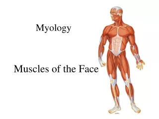
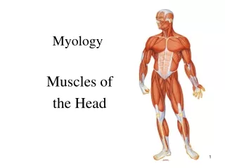
SPECIAL MYOLOGY Mm. capitis (Muscles of the head) • MUSCULI MASTICATORII (MASTICATORY MUSCLES) Innervation - n. trigeminus = V. cranial nerve 2) MUSCULI FACIALES (MUSCLES of FACIAL EXPRESSION) Innervation - n. facialis = VII. cranial nerve NO FASCIA! – skin muscles
Mm. faciales(mimic muscles)(facial nerve – n.VII.) Muscles of the scalp Muscles of the orbit region Muscles of the nasal region Muscles of the mouth region Their contraction causes shift of the skin (folds or wrinkles) – it is the basis of the facial expression. They have no fascias!
Muscles of the neck (mm. colli) Superficial layer m. platysma m. sternocleidomastoideus mm. suprahyoidei(depression of mandible) mm. infrahyoidei - mainly fixation of os hyoideum (hyoid bone) Deep layer - mainly flexion of the neck (and head) mm. scaleni mm. prae- and intervertebrales
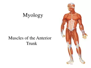
Musculithoracis, abdominis et dorsi (Musclesofthechest, abdomen and back)
Musculithoracis(thoracicmuscles) 1. Thoracohumeralmuscles mainlyventralflexion and abduction oftheupper limb Musculus pectoralis major Musculus pectoralis minor Musculus subclavius Musculus serratus anterior 2. True (original) thoracic muscles muscles for respiratory movements Musculi intercostales externi, interni et intimi Musculus transversus thoracis 3. Diaphragma mainmuscle for inspiration
Musculiabdominis(abdominalmuscles) antagonistsofthedorsalmuscles, regulatethevolumeoftheabdominalcavity
Musculiabdominis(musclesofthe abdomen) antagonistsofthedorsalmuscles, regulatethevolumeoftheabdominalcavity Ventral group musculus rectus abdomis (+ its sheat=vagina mm. recti abdominis) musculus pyramidalis Lateral group musculus obliquus externus abdominis musculus obliquus internus abdominis musculus transversus abdominis musculus cremaster Dorsal group musculus quadratus lumborum Canalis inguinalis (inguinal canal)!!!
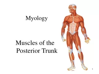
Musculi dorsi (muscles of the back) I. Extrinsicmusclesoftheback II. Intrinsicmusclesoftheback(locateddeeper, innervation by dorsalramiofspinalnerves) III. Shortmusclesoftheback Ad I. Extrinsicmusclesoftheback A) Mm. spinohumerales(spinohumeralgroup)movementsoftheupper limb B) Mm. spinocostales(spinocostalgroup)helprespiratorymovements
I. EXTRINSIC BACK MUSCLES A. Mm. spinohumerales (spinohumeral group) 1. m. trapezius 2. m. latissimus dorsi 3. m. levator scapulae 4. m. rhomboideus minor 5. m. rhomboideus major
B) Spinocostal group of muscles help respiratory movements 1. m. serratus posterior superior 2. m. serratus posterior inferior
Ad II. Intrinsicmusclesoftheback mainlyextensorsoftheback and thehead, innervationramidorsalesofspinalnerves 1)Spinotransversalsystem(m. spleniuscapitis and cervicis) 2) Sacrospinalsystem(m. erectorspinae, longissimus and iliocostalis) 3) Spinospinalsystem(m. spinalisthoracis) 4) Transversospinalsystem(m. semispinaliscapitis and cervicis)
Ad III. Short muscles of the back Mm. nuchae profundi a)m.rectus capitis posterior minor(lesser) b) m. rectus capitis posterior major (greater) c) m. obliquus capitis superior d) m. obliquus capitis inferior Trigonum suboccipitale (bordered by b, c, d) Content: a. vertebralis a. cervicalis profunda n. suboccipitalis dorsal arch of the atlas 2 1
Fasciae of dorsal muscles Fascia dorsi superficialis Fascia nuchae Fascia thoracolumbalis (actually aponeurosis of m. latissimus dorsi – its lamina superficialis) Aponeurosis lumbalis (lamina profunda of fascia thoracolumbalis, separates m. quadratus lumborum from m. erector spinae)
Used pictures come from: Moore, K. L. (1992): Clinical oriented anatomy. Third edition. Williams&Wilkins, A Waverly Company. Gilroy, A. M. et all. (2009): Atlas of Anatomy. Thieme New York, Stuttgart. Putz, R. (2008): Atlas of Human Anatomy Sobotta. Elsevier Books. Platzer, W., Kahle, W., Leonhardt H. (1992): Locomotor system. Georg Thieme Verlag, Stuttgart, New York, 4th edition. Čihák, R. (1987): Anatomie 1. Avicenum, Zdravotnické nakladatelství.