Download
1 / 54
990 likes | 1.76k Vues
BI-RADS, C-RADS, CAD-RADS, LI-RADS , Lung-RADS , NI-RADS, O-RADS, PI-RADS , TI-RADS: Reporting and Data Systems. Julie Y. An , MD Kyle M.L. Unsdorfer , MD Jeffrey C. Weinreb , MD.

E N D
BI-RADS, C-RADS, CAD-RADS, LI-RADS, Lung-RADS, NI-RADS, O-RADS, PI-RADS, TI-RADS: Reporting and Data Systems Julie Y. An, MD Kyle M.L. Unsdorfer, MD Jeffrey C. Weinreb, MD An earlier incorrect version of this presentation appeared online. This presentation was corrected on September 12, 2019.
Author Affiliations From the Department of Medicine, Akron, Ohio 44304 (J.Y.A.); Department of Radiology, Mayo Clinic, Rochester, Minn (K.M.L.U.); and Department of Radiology, Yale University School of Medicine, New Haven, Conn (J.C.W.). Address correspondence to: J.Y.A. Current address: Department of Radiology, University of California San Diego 200 W Arbor Drive, San Diego, CA 92103 (e-mail: j1an@ucsd.edu) 2018 RSNA Educational Exhibit:MS101 Acknowledgments.—The authors would like to thank Philip Araoz, MD, Chi Wan Koo, MD, and Baris Turkbey, MD for their guidance and review of this presentation. Disclosures of Conflicts of Interest.—J.C.W. Activities related to the present article: member of the ACR LI-RADS and PI-RADS committees; received reimbursement for travel expenses for committee meetings from the ACR. Activities not related to the present article: disclosed no relevant relationships. Other activities:disclosed no relevant relationships.
Review the overview of the current American College of Radiology (______)* Reporting and Data Systems (RADS). Recognize the systematic scoring and modifiers of RADS. Review the general themes and key imaging components of each system. Identify additional resources for more in-depth training and education. Note.—*Reprinted, with permission, from the ACR.
Why Standardized Reporting? • Unifies the language between radiologists and clinicians • Allows consistent data collection and improvement of diagnostic parameters • Helps to standardize care • Adds value by providing clinical recommendations and guidance
What is ______RADS? • RADS include systems with standardized terminology, assessment, and reporting endorsed by the ACR. • The predominant focus is on cancer imaging (breast, colon, head and neck, liver, lung, ovarian, prostate, and thyroid cancers), with additional systems focusing on coronary artery disease and head injuries (pending publication). • Systems were devised by expert consensus and may be updated periodically.
“The goal of the________ RADS is to reduce the variability of terminology in reports and to ease communication between radiologists and referring physicians.”1
ACR RADS Abbreviations BI-RADS= Breast Imaging Reporting and Data System C-RADS= CT Colonography Reporting and Data System CAD-RADS= Coronary Artery Disease Reporting and Data System LI-RADS= Liver Imaging Reporting and Data System NI-RADS = Neck Imaging Reporting and Data System O-RADS= Ovarian-Adnexal Reporting and Data System PI-RADS= Prostate Imaging Reporting and Data SystemTI-RADS=Thyroid Imaging Reporting and Data System
TI-RADS NI-RADS Thyroid cancer Head and neck cancer Lung-RADS BI-RADS Lung cancer Breast cancer LI-RADS Hepatocellular carcinoma CAD-RADS Coronary artery disease C-RADS Colon cancer O-RADS PI-RADS Ovarian and adnexal mass Prostate cancer
*Cancer Specific TI-RADS NI-RADS Thyroid cancer Head and neck cancer Lung-RADS BI-RADS Lung cancer Breast cancer LI-RADS Hepatocellular carcinoma CAD-RADS Coronary artery disease C-RADS Colon cancer O-RADS PI-RADS Ovarian and adnexal mass Prostate cancer
_______Reporting and Data Systems Note.—C = prior lung cancer, CEUS = contrast material–enhanced US, HCC = hepatocellular carcinoma, M = malignant but not HCC, NA = not applicable, N, NC = inadequate study, S = clinically significant or potentially clinically significant findings (nonlung cancer), TIV = tumor in vein, TR = treated.
RADS Scoring Scale 1____2____3____4____5____6 Increased likelihood of disease
RADS Scoring Scale Increased likelihood of disease 1 2 3 4 5 C1 C2 C3 C4 1 2 3 4 5 1 2 3 4 5 1 2 3 4A 4B 4X 1 2A 2B 3 4 1 2 3 4 5 1 2 3 4 5
Nondiagnostic Scores 0,N,NC Inadequate study, requires repeat or additional imaging *CAD-RADS 0 =Exception, not an inadequate study
Subscores 4A - Low suspicion 4B - Intermediate suspicion 4C -Moderate suspicion 4A - Smaller, less solid 4B - Larger, more solid 4X -Has additional findings that raise suspicion 2A - Superficial lesion 2B - Deep lesion
Score Modifiers C - Colonic findings E - Extracolonic findings M - Malignant but not HCC TR - Treated TIV - Tumor in vein C - Prior lung cancer S - Significant findings, not lung cancer
Imaging Examples of ______RADS1
BI-RADS2 • BI-RADS is appropriate to use for patients undergoing screening, diagnostic, or posttreatment follow-up imaging for breast cancer. • First system proposed in 1993, and the first RADS to gain widespread clinical adoption • Integrated with the American Society of Breast Surgeons guidelines Pathologic condition: Breast cancer (screening and diagnostic) Imaging modalities: Mammography, MRI, US Scoring: Patient score 0–6 (4A-C) Version: 5 (2015)
BI-RADS • Additional Categories • BI-RADS 0:Incomplete assessment Need to perform repeat imaging or to review prior mammograms for comparison • BI-RADS 6: Known biopsy-proven malignancy Appropriate action should be taken
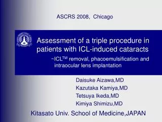
C-RADS3 • C-RADS is used for patients undergoing screening CT colonography. • From the Working Group on Virtual Colonoscopy, which includes members of the ACR Colon Cancer Committee • Reports colorectal neoplasia and extracolonic findings Pathologic condition: Colon cancer Imaging modality: CT colonography Scoring: Lesion score C0–4, E0–4 Version: 1 (2005)
C-RADS Retroaortic left renal vein Simple renal cyst Minimally complex renal cyst Portal thrombus and metastasis Additional Categories.—C0: Inadequate study: inadequate preparation, insufflation, awaiting prior studies for comparison Repeat examination and review prior studies E0: Limited examination: artifact, evaluation of extracolonic soft tissue is severely limited Alternative workup indicated
CAD-RADS4 • CAD-RADS is used for patients with stable or acute chest pain. Additional inclusion criteria: • Negative first troponin test results • Negative or nondiagnostic electrocardiogram (EKG) • Thrombolysis in myocardial infarction (TIMI) score less than 4 (low-intermediate mortality risk of unstable angina/non–ST-segment elevation myocardial infarction [NSTEMI]) • Graded by the patient’s highest- risk coronary lesion Pathologic condition: Coronary artery disease Imaging modality: Coronary CT angiography Scoring: Patient score 0–5, N Version: 1 (2016)
CAD-RADS Normal LAD >50% narrowing proximal to a stent. CAD-RADS 3S(S = stent) Noncalcified plaque in mid LAD without stenosis Severe 80%–90% stenosis Proximal left main calcified plaque leading to a 25% stenosis Additional Category.—CAD-RADS N – Nondiagnostic study. CAD cannot be excluded. ACS cannot be excluded Additional and/or alternative evaluation for ACS needed Note.—ACS = acute coronary syndrome, ICA = invasive coronary angiography, LAD = left anterior descending coronary artery.
LI-RADS5–7 • LI-RADS applies to patients at high risk for HCC (eg, those patients with hepatitis B or C, cirrhosis from nonalcoholic steatohepatitis [NASH], alcohol, or prior HCC • Excludes patients younger than 18 years of age, those with no risk factors and/or cirrhosis, vasculopathies, or Budd-Chiari syndrome • Integrated with the American Association for the Study of Liver Diseases (AASLD) guidelines Pathologic condition: HCC Imaging modalities: CT, MRI, US, contrast-enhanced US Scoring: CT, MRI, contrast-enhanced US = lesion score, 1–5, TIV, NC, M, T US = Patient score,1–3 Version: 3 (2017–2018)
LI-RADSCT/MRI • Note.—LR-5 threshold growth = 50% diameter increase over 6 months. • Reprinted, with permission, from reference 5.
LI-RADSCT/MRI • Additional Diagnostic Categories • LR TIV - TIV Multidisciplinary discussion, may need to perform a biopsy • LR NC – Nondiagnostic owing to image degradation or omission Repeat or perform alternative imaging in 3 months or less • LR M - Probably malignant, not necessarily HCC Multidisciplinary discussion, may need to perform a biopsy • Additional Treatment Response (TR) Categories • TR Nonevaluable- Inadequate imaging technique and/or quality • TR Nonviable - Low likelihood of viable tumor • TR Nonviable - Moderate likelihood of viable tumor • TR Equivocal - High or definite likelihood of viable tumor PVP Delayed 7 mm. No APHE, WO, or capsule. Definitely a cyst. Definitely hemangioma PVP PVP 8 mm with APHE. No WO or capsule. Possible cavernous hemangioma. Probably hemangioma PVP PVP 6 mm with APHE and WO. No capsule. Possible HCC. 10 mm with APHE. No WO or capsule. Possible HCC. PVP Delayed 14 mm with APHE and WO. No capsule. Probably HCC. 17 mm with APHE and WO. No capsule. Probably HCC. Note.—APHE = arterial phase hyperenhancement, FNH = focal nodular hyperplasia, HC = hepatocellular, PVP = portal venous phase, WO = washout. Delayed Delayed PVP 16 mm with APHE, WO, and capsule. HCC. 23 mm with APHE, WO, and capsule. HCC.
LI-RADSUS 9-mm hyperechoic observation in superior left lobe 17-mm heterogeneous mass in segment VIII
Lung-RADS8 • Lung-RADS is used for screening examinations of adults ages 55–80 (per the U.S. Preventive Services Task Force) with 30 pack-year (pkyr) smoking history and those who quit within 15 years. • Recommended annual repeat screening • Patient level score determined based on the nodule with the highest degree of suspicion Pathologic condition: Lung cancer (screening) Imaging modality: Low-dose CT Scoring: Patient score 0–4 (4A,B,X), S, C Version: 1 (2014)
Lung-RADS Part-solid 5-mm nodule Solid 7-mm nodule 4X solid ≥ 15-mm nodule with spiculation Additional Categories: Lung-RADS 0 - Incomplete: Prior chest CT examinations being located for comparison, part or all of lungs cannot be evaluated Lung-RADS S - Other: Clinically significant or potentially clinically significant findings that are nonlung cancer Lung-RADS C - Prior lung cancer: Patient with prior diagnosis who is returning for screening
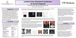
NI-RADS9 • NI-RADS is used for patients with a new neck mass and those with a known and/or treated mass undergoing surveillance • Provides recommendations regarding surveillance, direct inspection, short-term follow-up, additional imaging, and performing biopsy. Pathologic condition: Head and neck cancers (diagnostic and surveillance system) Imaging modalities: PET, CT, MRI Scoring: Patient score 0–4 (2A-B) Version: 1 (2016)
NI-RADS • All images reprinted, with permission, from reference 9. Additional Categories NI-RADS 0 – Incomplete: New baseline study and knowledge of prior imaging exists and will be available for interpretation Assign score in addendum after prior imaging becomes available
O-RADS10 • O-RADS is a standardized lexicon, not a scoring system. • Discourages the use of colloquial terms such as complex, ring of fire, etc • Evaluation by laterality • Pending expansion of the lexicon for MRI Pathologic condition: Ovarian and adnexal masses Imaging modality:Transvaginal US Scoring: Not scored Version: 1 (2018)
O-RADS Color score 2 – minimal flow Color score 1 – no blood flow Color score 4 – marked flow Color score 3 – moderate flow Note.—IOTA = International Ovarian Tumor Analysis.
PI-RADS uses multiparametric MRI sequences, T2-weighted imaging, diffusion-weighted imaging (DWI), apparent diffusion coefficient (ADC) mapping, and dynamic contrast-enhanced (DCE) imaging, for the detection, surveillance, and biopsy planning of prostate lesions. • Dominant sequences performed for scoring is based on the lesion location: PI-RADS11 Pathologic condition: Prostate cancer Imaging modality: Multiparametric MRI Scoring: Lesion score 1–5 Version: 2 (2015) D T Peripheral Zone (PZ) = DWI Transition Zone (TZ) = T2-weighted imaging • Artwork reprinted and adapted, with permission, from reference 11.
PI-RADS Right mid-PZ: DCE score = negative Right-mid PZ: DWI score = 2 Right-mid PZ: DWI Score = 3 Right-mid PZ: DCE = negative Right Apical-mid PZ: DWI score = 3 Right Apical-mid PZ: DCE = positive Left TZ: T2 score = 5 Left TZ: DWI score = 5
TI-RADS12 • TI-RADS is used for risk stratification in patients with thyroid nodules and offers guidance on fine-needle aspiration (FNA) and follow-up • Scored by the summation of points given to imaging characteristics suggestive of malignancy • Free online calculators are available. Pathologic condition: Thyroid cancer Imaging modality: US Scoring: Lesion score 1–5 Version: 1 (2017)
TI-RADS Sum points from each category
Current Limitations • Adoption varies from institution to institution, as does experience among radiologists • Lack of consistency in terminology and structure. For example: • lesion or observation • category, score, or level • category ranges: 0,1–4, 5, 6 • Nondiagnostic findings represented by 0, N, or NC • May be improved by • Combining oncology RADS to five main types to avoid confusion. For example: • 1 = benign, 2 = likely benign, 3 = indeterminate, 4 = likely malignant, 5 = malignant • Standardizing the terminology used for nondiagnostic and negative studies • Limiting RADS to cancer imaging
Future Direction • ACR is currently changing the organization and approval process for RADS efforts in order to deal with growing demands. • Additional RADS are in varying stages of development. • The Myeloma Response Assessment and Diagnosis System (MY-RADS)13 (whole-body MRI for myeloma), Vesical Imaging-Reporting and Data System (VI-RADS)14 (MRI for bladder cancer), Metastasis Reporting and Data System for Prostate Cancer (MET-RADS)15 (whole-body MRI for metastatic prostate cancer) are recent non–ACR endorsed systems. • Incorporation of standardized templates and terminology with vendor-provided ACRassist • Integration with clinical society guidelines to increase adoption • For example, LI-RADS with the AASLD and BI-RADS with the American Society of Breast Surgeons guidelines
Publication Trends • BI-RADS and PI-RADS have had the greatest number of publications. • Suggests these systems have the greatest clinical and research interest Data obtained from reference 16. Note: BI-RADS and PI-RADS publication frequency ranges from 0 to 300 articles vs 0 to 30 articles for all other systems.
Publication Trends • Growing number of articles published on LI-RADS, TI-RADS, Lung-RADS, and CAD-RADS Data obtained from reference 16. Note: BI-RADS and PI-RADS publication frequency ranges from 0 to 300 articles vs 0 to 30 articles for all other systems.
Publication Trends • C-RADS, O-RADS, and NI-RADS have not had as much research interest. • Notably, these three systems have not been updated since their initial release Data obtained from reference 16. Note: BI-RADS and PI-RADS publication frequency ranges from 0 to 300 articles vs 0 to 30 articles for all other systems.
______Website • Free, detailed, and up-to-date resources for new systems and recent changes1 • Ex: Head Injury Imaging Reporting and Data System (HI-RADS) is currently being developed for imaging traumatic brain injury • Reprinted, with permission, from reference 1.
TI-RADS Video Resources Three-part ACR TI-RADS Webinar ACR TI-RADS Steering Committeehttps://www.youtube.com/watch?v=s0f1cU7rxXA&t=113s17 https://www.youtube.com/watch?v=Y9JU2i4IF-M&t=111s18 https://www.youtube.com/watch?v=fsWAle6u-uE&t=333s19 ACR TI-RADS SeriesJenny Hoang, MDhttps://www.youtube.com/watch?v=lAeyb-QrXm4&t=7s 20 https://www.youtube.com/watch?v=P7GHtG_9GvQ 21 https://www.youtube.com/watch?v=w4oZYkw8wfw&t=2s 22 Images reprinted, with permission, from references 20–22. Images reprinted, with permission, from references 17–19. LI-RADS Introduction to LI-RADS, Claude Sirlin, MD https://www.youtube.com/watch?v=Fml7wH5ps90&t=1s 23 2017 Version of LI-RADS for CT and MR Imaging: An Update RadioGraphicshttps://www.youtube.com/watch?v=q-4BEMvhrNg 24 Image reprinted, with permission, from reference 23.
Video Resources BI-RADS Breast Cancer Review Series: BI-RADS 5th Edition Leonardo Valentin, MD https://www.youtube.com/watch?v=AYZcnhRwdxo&t=1s25 Image reprinted, with permission, from reference 25. Prostate MRI: Common Imaging Pitfalls Art Rastinehad, DOhttps://www.youtube.com/watch?v=7L0VC9JY2ak27 PI-RADS Image reprinted, with permission, from reference 26. Prostate MRI Using PI-RADS Andrew Rosenkrantz, MD https://www.youtube.com/watch?v=GJPQ1a4xjAs&t=25s26 Image reprinted, with permission, from reference 27.
Interactive Cases Lung-RADS Lung Cancer Screening eLearning Program (ACR login required) https://www.acr.org/Lifelong-Learning-and-CME/Learning-Activities/Lung-Cancer-Screening-Education28 BI-RADS Image reprinted, with permission, from reference 28. Mammography Case Review (ACRMC7 forthcoming) https://www.acr.org/Lifelong-Learning-and-CME/Learning-Activities/Mammo-Case-Review29 LI-RADS Image reprinted, with permission, from reference 29. LI-RADS teaching and case-based module, Montefiore/Albert Einstein, (ACR login required) https://cortex.acr.org/Presenters/CaseScript/CaseView?CDId=bSaQzu9QeR0%3d30 PI-RADS Learn Prostate MRI Andrew Rosenkrantz, MD http://learnprostatemri.com/interactive-cases/31 Image reprinted, with permission, from reference 31. Image reprinted, with permission, from reference 30.
Summary • The ACR-endorsed RADS are a set of guidelines for the evaluation and interpretation of disease-oriented imaging studies. • Systems may be periodically updated to improve diagnostic parameters, and new systems are in various stages of development. • Clinical adoption of ACR RADS varies. • BI-RADS, followed by PI-RADS and LI-RADS, have the greatest acceptance. • Integration of RADS into multidisciplinary guidelines has the potential to increase the field of radiology’s impact on clinical care.