Rheumatology Labs
390 likes | 654 Vues
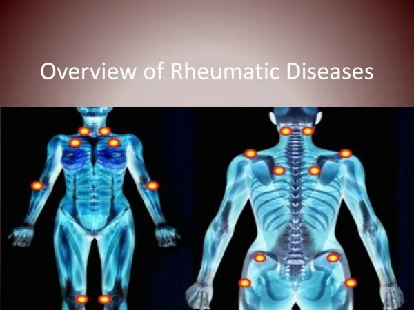
Rheumatology Labs. Brenda Beckett, PA-C Clinical Assessment II. Rheumatology. Evaluation of the patient with a suspected rheumatologic disorder is dependent upon an initial clinical suspicion, a thorough history and clinical evaluation, and laboratory testing. Collagen Vascular Disorders.

Rheumatology Labs
E N D
Presentation Transcript
Rheumatology Labs Brenda Beckett, PA-C Clinical Assessment II
Rheumatology Evaluation of the patient with a suspected rheumatologic disorder is dependent upon an initial clinical suspicion, a thorough history and clinical evaluation, and laboratory testing.
Collagen Vascular Disorders • Considerable overlap exists in the clinical features of these disorders as well as in the presence of the various antibodies. (nonspecific) • Systematic approach to the laboratory diagnosis will more often result in a correct diagnosis and in many instances shorten the time and decrease the expense incurred before appropriate therapy can be initiated.
Antinuclear Antibodies (ANAs) • Group of antibodies directed against proteins in cells. • ANA test is not specific to any one antibody (but it is sensitive to SLE) • Multiple follow-up tests to confirm positive
Antinuclear Antibody (ANA) • Initial screening test used in the evaluation of patients with a suspected collagen vascular disease (RF should also be tested for in adults with arthritis). • ELISA or other screen to rule out negatives. • Positives are tested by indirect immunofluorescence, have patterns that are easy to read.
Antinuclear Antibody (ANA) • Titer greater than or equal to 1:160 is considered a significant positive. • Titers less than or equal to 1:80 are usually of no or questionable significance. • Positive ANA incidence is 99% in SLE, 85% in Sjögren's, 88% in scleroderma, 55% in rheumatoid arthritis, and 40% in juvenile rheumatoid arthritis.
Antinuclear Antibody (ANA) The ANA consists of antibodies of different specificities. There is controversy about the usefulness of determining the pattern of immunofluorescence staining of the ANA and the relationship of the pattern to antibody specificity and disease state.
Generalizations Regarding ANA Patterns • Peripheral (Rim) Pattern of Staining • suggests anti-DNA antibodies, seen most often in SLE. • Homogeneous Staining • suggests anti-DNA, anti-histone, or anti-deoxyribonucleoprotein (DNP) antibodies observed in SLE, RA, and drug-induced SLE.
Generalizations Regarding ANA Patterns • Speckled Pattern • (1) Antibody to SSA or SSB, observed in SLE and SS • (2) Smith antibody (Sm) observed almost exclusively in SLE • or (3) RNP antibody observed in SLE, MCTD, RA, and scleroderma
Generalizations Regarding ANA Patterns • Anti-nucleolar Staining • observed in scleroderma and some forms of Raynaud's phenomenon. • Centromere Pattern • observed in the CREST syndrome of scleroderma • Calcinosis • Raynaud's phenomenon • Esophageal hypomotility • Sclerodactly • Telangiectasia
ENA • Extractable Nuclear Antigen Antibodies. (RNP – Ribonucleic Protein, Smith, Scleroderma, SSA and SSB – Sjögren’s Antibodies). • Antibodies to nuclear antigens are hallmarks of collagen vascular diseases
ENA • Sm - Specific for SLE • SSA (Ro)- SLE, Sjögren’s • SSB (La)- SLE, Sjögren’s • RNP - SLE, Sjögren’s, Scleroderma • Scl-70 - Scleroderma, some Sjögren’s • Jo-1 - Polymyositis/dermatomyositis
dsDNA Double stranded DNA antibody • High titers in SLE • RA, Sjögren’s • ssDNA test also positive in SLE, but not as specific
Rheumatoid Arthritis (RA) • Immunologic expression of an individual's immune system reaction to the presence of an immunoglobulin molecule that is recognized as "non-self." • This response to the "non-self" immunoglobulin results in the presence of immune complexes. These, in turn, bind complement and may eventually lead to synovium, cartilage, and bone destruction
Rheumatoid Factor (RF) • RFs are immunoglobulins directed against portion of IgG • RF may be found in a variety of autoimmune diseases, as well as in up to 10% of apparently healthy individuals. • May be positive in some patients with syphilis, viral infections, chronic liver diseases, sarcoidosis, leprosy, neoplasms, and a variety of other chronic inflammatory conditions (nonspecific)
Rheumatoid Factor • High concentrations of RF (>300 IU/mL) are found with RA and Sjögren's syndrome. • Juvenile onset RA is less likely to have a positive test for RF. • The RF test should be used with caution in the diagnosis of RA because of the low predictive value of the test for this disease. The percentage of positive RF assays in the normal population increases with age.
Anti-CCP • Anti-cyclic citrullinated peptide (immune protein) • As sensitive as, and more specific than RF in early and fully established disease • May predict RA in undifferentiated arthritis • A marker of erosive disease in RA • May be detected in healthy individuals years before onset of RA
Antineutrophilic Cytoplasmic Antibodies (ANCAs) • Important for the diagnosis of certain vasculitis syndromes (Wegener’s granulomatis) • cANCA (cytoplasmic) • pANCA (perinuclear) • Also positive in RA, SLE, myositis, infections, GI disease, etc
HLA B27 • Human Leukocyte Antigen B27 • 90% of patients with Ankylosing Spondylitis have HLA B27 • Only small amount of patients with HLA B27 will have disease • Can also be at risk for Reiter’s, IBD, Psoriatic Arthritis
C-reactive Protein (CRP) Protein that binds to polysaccharides present in many bacteria, fungi, and protozoal parasites. It also binds to phosphocholine, lecithins, and polyanions such as nucleic acids. Once complexed, CRP becomes an activator of the classical complement pathway. CRP recognizes potentially toxic autogenous substances released from damaged tissues, binds them, and then detoxifies or clears them from the blood.
C-reactive Protein (CRP) CRP is the most sensitive acute phase protein. CRP will elevate within two hours of acute insult (surgery, infection, etc.) and should peak and begin decreasing within 48 hours if no other inflammatory event occurs.
C-reactive Protein (CRP) • Dramatic increases following MI, trauma, infection, surgery, or neoplastic proliferation. • In patients with rheumatoid arthritis, persistently elevated CRP concentrations are present when the disease is active and usually fall to normal during periods of complete remission.
Erythrocyte Sedimentation Rate (ESR) • Nonspecific marker of inflammation. Generally, ESR does not change as rapidly as does CRP, either at the beginning of inflammation or as it resolves. CRP is not affected by as many other factors as is ESR, making it a better marker of inflammation. Many clinicians still use ESR as an initial test due to its wide availability and low cost.
Synovial Fluid • Lubricates cartilage in joint space • Normal: • Small amount • Clear, acellular (low WBC count) • High viscosity • Does not clot • Protein = 1/3 plasma protein • Glucose approximates plasma glucose
Synovial Fluid • Detect inflammation, infection • Crystals • Monosodium urate – gout • Calcium pyrophosphate - pseudogout
Gout • Synovial fluid analysis – to detect monosodium urate crystals. • Serum Uric Acid may be elevated • Urine Uric Acid may be elevated • Kidney function tests – to determine if uric acid crystals are accumulating in the kidneys and impairing their function.
Case Study History and Presentation • 60 yo female admitted cc of generalized weakness x2wks, morning stiffness both knees, aggravated by movement. Mild fever. Lost 10lbs in one mo. No significant medical hx, except similar episodes 2x in past year, involving hands and wrists - took NSAIDS.
Case Study Physical Exam • A&O, mild distress. BP 120/80, HR 85, T 1000F, RR 20. Abd soft, nontender, mild splenomegaly. Knees swollen bilat, warm, painful with movement. Mild atrophy of forearm muscles with small subcu firm nodules in posterior forearms. Swelling of MCP joints.
Case Study • X-ray of hand taken: • Showed periarticular osteoporosis, bone and cartilage destruction, joint space narrowing.
Case Study • Differential Diagnosis?
Case Study • What lab tests do you want to order?
Case Study • What is your diagnosis? - • What other test would you consider performing to verify your diagnosis? - • What else should we follow-up on in this patient? -