Download

1 / 34
380 likes | 2.48k Vues
Burns Fluid & Electrolytes. Nursing Care BURN INJURIES. 1. Identify the mechanism of burn (TYPE) injuries. 2. Describe methods for determining a ssessment /physiology/ classification of burns.

E N D
Nursing Care BURN INJURIES 1. Identify the mechanism of burn (TYPE) injuries. 2. Describe methods for determining assessment/physiology/ classification of burns. 3. Differentiate degrees of burn (1st- 4th) versus epidermal/superficial, partial and full thickness, deep burns. 4. Determine nursing care based upon the systemic pathological changes associated with burn injury in the first 24 – 48 hours. 5. Identify assessment, nursing diagnoses and management of the burn victim’s airway, breathing and circulation and wounds. 6. Identify the Pain/Nutritional/Rehab requirements for a burns patient.
Mechanism/Burn Type • Thermal - burning of tissue via direct contact with a heat source hot water, flame • Zones of Injury • 1. Zone of coagulation – thrombosis, vasoconstriction, necrosis and cell death • 2. Zone of stasis - low blood flow • 3. Zone of hyperemia - inflammatory response
Mechanism/Burn Type • Chemical - tissue destruction via direct contact chemical · oxidizing agent: sodium hypo chloride · reducing agent : hydrochloric acid · corrosives : phosphorus · protoplasmic poisons : formic acid · desiccants : sulfuric acid · vesicants : mustard gas · gasoline
Mechanism/Type:ElectricalBurn • - direct contact with electrical current ® entry & exit wounds
Burns Assessment/Physiology/ Classification Based on: • Depth/Degree of injury, • Percent of body surface areas involved, • Location of the burn, • Association with other injuries.
Burns Physiology/Classification Depth of Burn Assessment Epidermal : destruction epidermis only · reddened, · blanches to pressure, · no blisters · painful · healing 3-5 days · no scarring
Burns Physiology & Classification Depth/Degree of Injury • First Degree: superficial, epidermal damage • erythematous & painful due to intact nerve endings • heal in 5-10 days • pain resolves within 3 days • no residual scarring
Burns Physiology & Classification Depth/Degree of Injury • Second Degree: partial thickness, epidermis/ dermis • superficial burns – moist, blister; • deeper burns - white and dry, blanch with pressure, and have reduced pain • heal in 10-14 days • can develop into third degree burns with infection, edema, inflammation and ischemia • treatment varies with degree of involvement - grafting is indicated for deep burns
Burns Physiology / Classification Depth of Burn Assessment Partial Thickness Superficial destruction epidermis to upper dermis ·bright red to pale ivory, blistered or weeping, blanches to pressure ·sensitive to pain, pressure temperature ·healing 14-21 days , no scarring
Burns Physiology/Classification Depth of Burn Assessment Partial Thickness deep destruction epidermis to deep dermis · mottled · white & waxy · blistering · diminished sensation to light pressure · healing months-weeks/usually scarring
Burns Physiology & Classification Depth/Degree of Injury • Third Degree: full-thickness, most severe of burns • results in necrosis and avascular areas • tough, waxy, brownish leathery surface with eschar, numb to touch • grafting required • usually have permanent impairment
Burns Physiology & Classification Depth/Degree of Injury • Fourth Degree: • full-thickness as well as adjacent structures such as fat, fascia, muscle or bone • reconstructive surgery is indicated • severe disfigurement is common
Burns Physiology & Classification Depth/Degree of Injury Full - destruction to epidermis, dermis, subcutaneous · dry, · pearly/yellow-charred, · does not blanch, · leathery, inelastic · minimal to no sensation of pain, healing via secondary granulation/graft
Burn Assessment Body Surface Area Rule of Nines • adult: 9% head; 9 % arms; 18 % legs ; 18 % chest; 18% back; 1 % perineum • child: 18% head; 9% arms; 14 % legs; 18 % chest; 18 % back
Burn Assessment • Location: • Important for assessing potential disability • greatest risk with face, eyes, ears, feet, perineum and hands • Upper extremities involved in 71% of burns, head and neck 52% • Associated Injuries: • Smoke inhalation • hoarseness, cough, singed nasal hairs, oral burns, wheezing • Carbon monoxide poisoning • Fractures • Trauma
Hospitalization in Major Burns · >10% surface area in children, elderly · >15% surface area in adults · specific regions - respiratory tract, face, neck, circumferential burns, hands, feet, major joints, genitalia, electrical burns, lightening burns · 3rd degree burns >3% children, >5% adults
Mortalityin Burns · >65% body surface area (BSA) · associated smoke inhalation · infection • >20% BSA with shock and other complications/related sequelae
Collaborative Nursing & Medical Management Pathology of the First 24 hours: · Temperature loss ® hypothermia · Plasma & Protein Loss · Hypovolemia/hemoglobin concentration · Tissue/blood destruction hypoxia · Release hemoglobin pigment/myoglobin ® ¯ GFR & UO · Tissue hypoxia and reduced renal function ® metabolic acidosis · Platelet destruction & of activation clotting cascade via intrinsic/extrinsic pathway ® DIC
Collaborative Nursing & Medical Management Pathology of the Second 48 hours: • temperature 2. fluid mobilization to intravascular space 3. renal loss K+ 4 Fluid resuscitation ® Serum Na+ ® dilutional coagulopathy
Collaborative Nursing & Medical Management Wound Care · tetanus toxoid > 50% BSA burn · and/or tetanus immunization ·chemical burns • irrigate all burns, cover until initial resuscitation complete ·electrical burns • AC current ® Tetany & risk Vent Fib High energy ® check # volts & blunt injuries
Collaborative Burn Management Primary Assessment & Resuscitation Airway check risks – event in an enclosed area, singed eyebrows/nasal hair, hoarse voice, stridor, wheeze, air entry/edema Breathing check risks – event in an enclosed area® evaluate for CO2 poisoning high PaO2 low SataO2
Collaborative Burn Management Circulation :Assessment & Resuscitation Parkland Formula – one of the most commonly used:First 24 hours an isotonic solution (Ringers Lactate)4mL/kg x TBSA% divide into 8 hour periods - first 50% in 8 hours - next 25% in 8 hours - final 25% in 8 hours urinary output should be 50-70mL/hr (1mL/kg) in the first 24 hours
Collaborative Burn Management Circulation Con’t: :Assessment & Resuscitation Second 24 hours Colloid/plasma is delivered 0.5mL/kg x TBSA for the next 8 hours. At 32 hours: 5% Dextrose + nutritional replacement require serial measurement serum electrolytes, urea, hematocit, blood albumin, urinary N+.
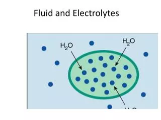
Nursing Diagnoses • Altered Tissue Perfusion • Fluid & Electrolyte Imbalance • Risk for Infection • Altered Comfort: Pain • Altered Nutritional: Less than Body Requirements (more Calories needed) • Body Image Change : Loss?: Role?
Nursing Care • IV access (Multiple) • Manage perfusion needs by parameters of CVP, Urinary Output • Pain management • once vital signs have stabilized, pain medication should be used (ie morphine, or meperidine, fentanyl, benzodiazepines as indicated ) • Morphine or Fentanyl Drip
Nursing Care of Ulcer/Pain/Tetanus • Curlings ulcer prophylaxis (Peptic Ulcer) • An H2 blocker (cimetidine, ranitidine,famotidine) start first 6 hours • antacids are no longer recommended - the patient should be kept NPO • with burns > 15% of BSA, an NG (OG) tube and bladder catheter should be placed • Tetanus • immunization if out of date
Nursing Care of Burn Wounds • Wound Care (Sterile Technique) • Debridement • Anti-microbial Application • silver sulfadiazine (Silvadine) • mafenide acetate (Sulfamylon) • Closed dressing except face & perineum • Wound cover • synthetic,biosynthetic, biological • Graft • Wound Allograft • Split thickness skin graft • full thickness graft
Evaluation of Nursing Care • ABC’s Airway – stridor • Breathing – use of accessory muscles, lung sounds • Circulation – CVP’s, BP, Pulse-Ox • Fluids & Electrolytes/Renal • Urinary output, labs, specific gravity, osmalarity, myoglobin • Pain • Infection (Gram Negative Sepsis) • Nutrition • Weight, ulcer Management