Download
1 / 85
1.33k likes | 4.48k Vues
Fetal Pig Dissection Review. GQ’s and Key Figures to Know. Introductory Website Activity. Online Exercise & Questions from the following website: http://bxscience.enschool.org/ourpages/auto/2009/2/5/65277921/Dissection%20of%20a%20Fetal%20Pig%20112.pdf

E N D
Fetal Pig Dissection Review GQ’s and Key Figures to Know
Introductory Website Activity • Online Exercise & Questions from the following website: http://bxscience.enschool.org/ourpages/auto/2009/2/5/65277921/Dissection%20of%20a%20Fetal%20Pig%20112.pdf **Do a search for TWO good websites that can be used during our dissection as reference. The site should have clear images that cover all the areas/systems we cover. Write these TWO sites down on our GQ #1
Orientation to the Guidebooks • Both are available on my website • Printing them out is optional
Dissection of the Fetal Pig - Introductory Preparation GQ #1 1. How many units are there in the manual? Which units have over six pages? 2. Using the anatomical directional terms written on page 4, determine which terms refer to... a)towards the head, b) towards the back, c) towards the toes, d) towards the middle. 3. What is the genus and species name for the domestic pig? What 2 characteristics are seen in all mammals? 4. A mammal that has 2 or more types of teeth has _______ dentition. 5. Pigs are considered to be even-toed ungulates. What other animals have similar settings? 6. What is the typical life span for a domestic pig? How many lbs. could the domestic pig weigh? How many offspring can a pig have? 7. How have pigs been useful subjects for humans other than for consumption (eating)?
General Rules • No Food or Drink at your Station at all times • Remain at your station at all times unless instructed • Keep track of all instruments (do a count everyday) • No open toed shoes • Do not cut anything unless instructed • Always follow your Dissection guide
Dissection Tasks • Researcher & Photographer (1-2 people): • Sets up computer (obtains images & info.) • Makes labels • Takes photographs, prints & shares with group • Answers all GQ’s and researches key information • Keeps the group in check with Dissection Manual • Equipment manager: • Cleans, dries, and returns all supplies to the appropriate places. • Cleans the table • Dissectors/Assistant( min. 2 people) - Observe the dissection procedures and share with rest of the group.
Materials Needed Daily • Computer w/website up and ready to use • Dissecting Manuals • Dissecting tray • Gloves • Paper towels • String • 3 Blunt probes • 4 Sharp Probes • 2 Scissors • 2 forceps • 12 Pins & Index Card (for labels later) • Large Zip-Lock Freezer Bag (Names, Period)
Unit 1 – External Features – Intro To Your Pig GQ #2 Name 3 important functions of the skin for your pig. What are the two layers of the skin? Name the four main sections of the pig. Another name for the nostrils are __________. What function does the nasal cavity serve? Does your pig have any teeth? Look inside and check. The third eyelid is called a(n)_______. What purpose does it serve? *Try to find this structure, you may need to make a small incision starting from the inner corner of the eye. Another name for the external ears is/are _____ and the openings is called the ______.
The trunk of your pig can be divided into the ______ and ______. • On the abdomen, what do you notice is present? What is the name for these structures? How many pairs are there? 10. What was the umbilical cord attached to? How far should you cut the umbilical from the abdomen? (Make the cut.) How many blood vessels do you see? How many of these are arteries? How many are veins? Does the blood from the fetus ever mix with the mother's? 11. Another name for the caudal opening of the digestive tract is _________________. 12. Following the directions on page 12, determine if your pig is a male or a female and be sure the correct name you've chosen is written on you bag. Males have a sac structure called a ________________ vs. females having _________________.
Unit 1 – External Features – Intro To Your Pig • Lab Partners’ names • Personal name of your pig • Take a close look at your pig, and give a boy’s and girl’s name, as you will determine the sex later. • Length of specimen ______ cm = ______ days * Measure from tip of nose to base of tail (See pg. 7) * Use piece of string and ruler
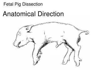
External Features to ID (Image Set #1) • Nares • Eyes & Nicitating membrane • Tongue • Pinnae • Thorax • Trunk • Umbilical cord (3 blood vessels) • Teats • Urogenital opening • Scrotum (male) or Genital papilla (female)-Pg. 9 • Anus • Measure length to estimate age of pig (use string and measure from the tip of the nose to the base of the tail) ****Use table on pg. 7 to estimate age.
External Features to ID (DAY 2) • Cut Umbilical cord approx. 2 cm from abdomen (pg. 8) • One Vein & Two Arteries should be see. • Nictitating membrane (3rd eyelid) • Make initial cuts through the skin using a scalpel. (See pg. 19) • Separate skin off specimen and begin isolating the muscles on ONE side of the pig by removing the fascia layer. • ID muscles indicated in the figures on Pgs. 20-22
GQ #3 - Unit 2 – The Skeleton • What does bone consist of? What makes bones hard? • What is the function of the bone? • Name the three types of joints. • Why can’t we use the fetal pig to study bones? • What part makes up the axial skeleton? • How many vertebral bones compose the cervical and thoracic areas. How do pigs differ with humans? • How many vertebral bones make up the lumbar and the sacral area in pigs? How do humans differ? • Name all the bones that make the cranium. • How many bones are facial? • How many ribs do pigs have? How do false ribs differ from floating ribs? • Name all the bones that make up the appendicular skeleton.
The Muscles Unit #3
GQ #4 • How many types of muscle tissue are there? Name them. Which types are voluntary and which ones are involuntary? • What muscles have striations? • Do muscles push or pull parts of the body? • When starting your dissection, what do you need the string for? • Your first incision needs to be SHALLOW and start at the ______ and continue up the _______. • Your second cut is along the ________. • The third cut is laterally across the _____ and along each____. • What do you need to do at the wrist? • How does your cut differ in male vs. female pig? 10. The following are responsible for what kinds of movements? Abductors, extensors, and constrictors. 11. The 2 adductor muscles of the shoulder are the pectoralis major and pectoralis minor. Identify them on your pig and ID/name the opposing abductors.
GQ #4 –Con’d 12. The triangular shaped muscle in the shoulder is called the ________. 13. The flexors of the upper arm include the ____ and ____. 14. The extensor of the upper arm is the _____. 15. In the neck, what are the functions of the following muscles? Masseter, Brachiocephalicus, Splenius (Identify and locate these muscles in your pig.) 16. Which muscles of the pig do we get bacon? 17. Which muscles do we get smoked ham from? (Fig. 14)
Muscle ID - Image Set #2 Ventral View, Shoulder, Neck & Upper arm Latissimus Dorsi Sternohyoid Triceps Deltoid Pectoralis Major Rectus Abdominus Trapezius Latissimus Dorsi Masseter Pectoralis Minor Extensor carpi radialis Extensor carpi ulnaris Brachioradialis Extensor digitorum lateralis Extensor communis Lat. View of Hind limb & Abdomen • Biceps Femoris • Gluteus maximus • Gluteus medius (medialis) • External Obliques • Semitendinosus
Exploration of the Neck (Image Set #3) • Sternohyoid Muscle • Larynx • Thyroid gland • Thymus gland • Trachea • Jugular vein • Carotid artery
The Respiratory System Units #4-5 Textbook See Chapter 19
GQ #5 - Unit 5 – Respiratory System • How many lobes are labeled in Fig.16 (p.27)? Name these lobes. • How does the trachea differ from the larynx? • What function does cilia and the rings of the cartilage serve? • List the following structures in order from the largest to the smallest: bronchioles, lungs, alveoli, bronchi, secondary bronchi • How many lobes are there for the right lung? Left lung? Why do you think there is a difference? • Locate/identify the pulmonary arteries & pulmonary veins. What color is each vessel? • What two cavities are separated by the diaphragm? • When the diaphragm contracts, is it moving up or down? Does this cause an inhaling or an exhaling reaction?
The human respiratory system Nasalcavity Pharynx (Esophagus) Left lung Larynx Trachea Rightlung Bronchus Bronchiole Diaphragm (Heart) Figure 22.6A
Larynx (upper part of respiratory tract) Vocal cords (sound production) Trachea (windpipe) Bronchi (tube to lungs) Bronchioles Alveoli (air sacs) Diaphragm (breathing muscle) Mammalian Respiratory Systems
Alveoli form the respiratory surface of the lungs • Oxygen diffuses through the thin walls of the alveoli into the blood • The bronchioles end in clusters of tiny sacs called alveoli Figure 22.6C Oxygen-richblood Oxygen-poorblood Bronchiole Alveoli Blood capillaries Figure 22.6B
Breathing • Negative pressure breathing: pulls air into lungs (mammals)-Vol. incr. • Inhalation: diaphragm contraction; Exhalation: diaphragm relaxation • Tidal volume: amount of air inhaled and exhaled with each breath (500ml) • Vital capacity: maximum tidal volume during forced breathing Regulation: CO2 concentration in blood (medulla oblongata)
This triggers a cascade of events Brain Cerebrospinal fluid • During exercise, the CO2 level in the blood rises, lowering the blood pH BREATHING CONTROLCENTERS—stimulated by: Pons Medulla CO2 increase / pH decreasein blood Nerve signalindicating lowO2 level Nerve signalstriggercontractionof muscles O2 sensorin artery Diaphragm Figure 22.9 Rib muscles
Smoking also causes emphysema • Cigarette smoke makes alveoli brittle, causing them to rupture • This reduces thelungs’ capacity for gas exchange • Smoking causes lung cancer and contributes to heart disease Figure 22.7A, B
It carries most of the oxygen in the blood • Hemoglobin is a protein in red blood cells Hemegroup Iron atom O2 loadedin lungs O2 O2 unloadedin tissues O2 Polypeptide chain Figure 22.10B
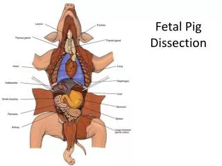
Exploration of the Thoracic Cavity (Image Set #4) • Lungs (7 lobes) • Diaphragm • Heart • Pericardium • Apex • Coronary artery & vein • Aortic arch • Atrium & Ventricles • Pulmonary vessels (arteries & veins) • Septum
GQ #5 - Unit 5 – Respiratory System • How many lobes are labeled in Fig.16 (p.27)? Name these lobes. • How does the trachea differ from the larynx? • What function does cilia and the rings of the cartilage serve? • List the following structures in order from the largest to the smallest: bronchioles, lungs, alveoli, bronchi, secondary bronchi • How many lobes are there for the right lung? Left lung? Why do you think there is a difference? • Locate/identify the pulmonary arteries & pulmonary veins. What color is each vessel? • What two cavities are separated by the diaphragm? • When the diaphragm contracts, is it moving up or down? Does this cause an inhaling or an exhaling reaction?
The Digestive System Unit 6 (See Textbook Chapter 17)
Lg. Intestine Cecum Ascending C. Transverse C. Descending C. Sigmoid C. Oral cavity Mouth Tongue Pharynx Salivaryglands Esophagus Liver Stomach Pyloricsphincter Stomach Gall-bladder Smallintestine Pancreas Smallintestine Sm. Intestine Duodenum Jejunum ileum Largeintestine Rectum Anus
Bile Liver Stomach Gall-bladder Acid chyme Bile Duodenum ofsmall intestine Pancreas Figure 21.10A
INTERIOR OF INTESTINE Blood vesselwith blooden route tothe liver Nutrientabsorption Nutrientabsorption Microvilli Epithelialcells Lumen Musclelayers Bloodcapillaries Circular folds Villi Lymphvessel EPITHELIALCELLS Nutrientabsorption VILLI INTESTINAL WALL Figure 21.10B
Identifying the Structures of the Digestive System(Image Set #5) • Hard palate • Soft palate • Esophagus • Liver (be able to name the 4 lobes) • Stomach (be able to identify cardiac region & pyloric region) • Spleen (Is it a digestive organ?) • Pancreas • Small intestine (Duodenum, Jejunum, & Ileum) • Large intestine –A, T, D, & Sigmoid (Fig.22 on p.35) • Cecum & Spiral colon *Isolate the small & Large intestines *Measure the length of the digestive tract Beg. Stomach to the anus
GQ #6 - Unit 6 – Digestive System • Name the five structures you need to identify in the pig’s mouth. (Fig.18) Make a cut on each side of the jaw to the mouth to open wider. See pg. 31 • What function does saliva serve? • Is the soft palate toward the front or the back of the mouth? • What purpose does the epiglottis serve? • How many lobes is the liver divided into? Name each lobe. • The veins of the liver are called the _____________ system. • What substance is stored in the gall bladder? Where is this substance made? What purpose does it serve? • Name the tubular structure that emerges from the gallbladder that serves to allow bile to travel to the small intestine. • Name the 3 functions of the liver. • What are the 2 substances (digestive juices) released in the stomach? • Name the circular muscle that prevents food from passing back up the esophagus. • What is the name of the green substance found inside your pig’s stomach? • What function does the pyloric sphincter serve? • What function does the pancreas and the spleen serve? Are they both part of the digestive system? • Why is the pancreas considered to be a “dual function” organ?
GQ #6 - Unit 6 – Digestive System • How long can the intestine be in your fetal pig? Name the divisions of the small intestine. Which one of the 3 segments is the shortest? Which is the longest? • “Material is prevented from passing prematurely into the large intestine (from the small intestine) by a sphincter known as the ________________________ valve. • What does the surface of the small intestine look like inside? What causes this appearance? • Name the three parts/regions of the large intestine. (not including the rectum and the anus, as mentioned in the dissection guide) • What purpose do the variety of bacteria play in the large intestine? (pg. 36)
Unit 7: Circulatory System (Pgs. 37-43) Textbook See Chapter 15
Guiding Questions #7 • Which side of the heart pumps blood to the pulmonary circuit? Do these vessels carry oxygen rich or oxygen poor blood? What is the name of the membrane that surrounds & covers the heart? • Blood that has been fully oxygenated leaves the heart through ____________ and transported to the rest of the body. Which side of the heart is the tricuspid atrioventricular valve (AV) located? How is this valve different from the mitral valve? • Arteries and veins that enter and leave the a) kidneys b)liver are called __________ arteries and _________ veins. • Venous blood enters the heart through the _________. • Name the arteries that supply the brain. Where are the iliac and femoral vessels located? • Trace the circulation pathway throughout the body starting at the right atrium. When is the blood oxygenated & deoxygenated? http://www.hillstrath.on.ca/moffatt/bio3a/fetalpig/frame01.htm (Very helpful pictures for the circulatory system – arteries and veins.)
Identifying the Structures of the Systemic Circuit (Image Set #6) • Locate & Identify the following Vessels: • Carotid Arteries, Jugular Veins • Subclavian & Brachial Arteries (upper extremities) • Abdominal Aorta & Vena Cava • Iliac & Femoral Arteries (lower Extremities) • Hepatic Arteries & Portal Veins • Renal Arteries & Veins • Mesenteric Arteries & Veins • Cross Section of the Heart: • ID Four Chambers • Coronary Arteries • Attempt to visualize the Four Valves • Pulmonary Arteries and Veins • Track the circulation of blood through the heart (Figs. 25,26, &27)
Circulation through the Heart • Superior & Inferior (Anterior & Posterior Vena Cava) • Right Atrium • Tricuspid Atrioventricular (AV) Valve • Right Ventricle • Pulmonary Semilunar Valve • Pulmonary Arteries • Lungs • Pulmonary Veins • Left Atrium • Bicuspid (Mitral) AV Valve • Left Ventricle • Aortic Semilunar Valve • Aortic Arch – Systemic Circuit