Download
1 / 9
90 likes | 258 Vues
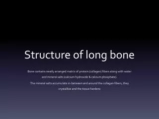
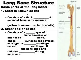
Anatomy of the Long Bone. The long bones are those that are longer than they are wide, and grow primarily by elongation of the diaphysis at an epiphysis at one end of the growing bone . The ends of epiphyses are covered with a hyaline cartilage ("articular cartilage").

E N D
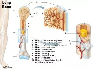
The long bones are those that are longer than they are wide, and grow primarily by elongation of the diaphysis at an epiphysis at one end of the growing bone. • The ends of epiphyses are covered with a hyaline cartilage ("articular cartilage"). • The longitudinal growth of long bones is a result of endochondral ossification at the epiphyseal plate. • Bone growth in length is stimulated by the production of growth hormone(GH), a secretion of the anterior lobe of the pituitary gland (it’s in your brain!). • The long bones include the femurs, tibias, and fibulas of the legs, the humeri, radii, and ulnas of the arms, and the phalanges of the fingers and toes.
The long bones of the leg comprise nearly half of adult height. • The other primary skeletal component of height is the spine and skull. • The outside of the bone consists of a layer of connective tissue called the periosteum. • Additionally, the outer shell of the long bone is compact bone, then a deeper layer of cancellous (spongy bone) which contains bone marrow. • The interior part of the long bone is the medullary cavity with the inner core of the bone cavity being composed of (in adults) of yellow marrow.
Articular Cartilage • Found on the ends of the bone • Allows smooth movement (articulation) within the joints and protects ends • There is no blood supply and it is insensitive • AKA- Hyaline cartilage Periosteum • The outer connective tissue covering the bone • Fibres, ligaments & tendons unite to connect bone to bone or muscle to bone • There is blood and nerve supply • It does not cover the articular cartilage
Medullary Cavity • The cavity of the diaphysis • Contains red (young) and yellow (adult) bone marrow • Red – blood cell formation • Yellow – adipose (fat) cell & connective tissue Compact Bone • Dense bone • Forms the walls of the diaphysis • Blood supplied via a system of canals • Can strengthen this part of the bone with exercise • Especially weight bearing that increase resistance and load
Cancellous Bone (Spongy) • Consists of an interwoven matrix of bone ( small cavity spaces) • These cavities are filled with marrow • They are called trabeculea • Continuous units of bony fibres arranged in a strut like system through cancellous tissue • exercise can strengthen this part of the bone just like compact bone
Epiphysis • The end of the long bone • Made up of cancellous bone covered with articular cartilage • Supplied by vessels from the joint capsule • Epiphyseal plate: growth plates, occur at various locations (Growth is possible) • Epiphyseal line: when the plates have fused and come together (cartilage no longer separates Epiphysis from Diaphysis) (Growth is no longer possible) Diaphysis • The central part of the long bone • Made up of the medullary cavity and surrounded by compact bone • Lined externally with the periosteum • Supplied by nutrient arteries