CARDIOVASCULAR FUNCTION
CARDIOVASCULAR FUNCTION . NUR-224. OBJECTIVES. Explain cardiac anatomy/physiology and the conduction system of the heart. Incorporate assessment of cardiac risk factors into the health history and physical assessment of the patient with cardiovascular disease.

CARDIOVASCULAR FUNCTION
E N D
Presentation Transcript
CARDIOVASCULAR FUNCTION NUR-224
OBJECTIVES • Explain cardiac anatomy/physiology and the conduction system of the heart. • Incorporate assessment of cardiac risk factors into the health history and physical assessment of the patient with cardiovascular disease. • Discuss clinical indications, patient preparation and other elated nursing implications fro common test and procedures used to assess and diagnose cardiovascular diseases.
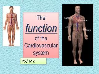
THE HEART Three layers • Endocardium • Myocardium • Epicardium • Four chambers • Heart valves
HEART • Surrounded by pericardium • Pericardial fluid 10-30 mL • Divided by septum • Left ventricular wall 2-3 x as thick as right ventricle • Atrial wall thinner than ventricles
BLOOD FLOW THROUGH THE HEART • Inferior and superior vena cava send deoxygenated blood to right atrium • Blood passes through tricuspid valve to right ventricle blood passes from right ventricle through pulmonic valve via pulmonary artery to lungs • Blood from lungs enters left atrium via pulmonary veins • Passes through mitral valve to left ventricle • Blood ejected to body through aortic valve aorta peripheral system
CONDUCTION SYSTEM • Depolarization (contraction of heart) • Sinoatrial node – pacemaker of heart • Contraction of atria • AV node • Bundle of His • Right and left bundle branches • Purkinje fibers
MECHANICAL SYSTEM Systole • Contraction of myocardium • Ejection of blood from ventricles Diastole • Relaxation of myocardium • Filling of coronary arteries • Atrium is emptying into the ventricles
HEART RATE • Number of times the ventricles contract each minute • 60-100 • Regulated by: Autonomic Nervous System Sympathetic Parasympathetic
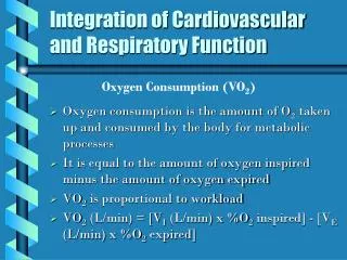
CARDIAC OUTPUT • Amount of blood pumped by each ventricle during a given period • Amount of blood ejected from ventricle with each beat (stroke volume) x heart rateCO = SV x HR 4 – 7 L/min
CARDIAC OUTPUT • Stroke volume: amount of blood ejected with each heartbeat • Cardiac output: amount of blood pumped by ventricle in liters per minute • Preload: degree of cardiac muscle fiber tension at end of diastole (prior to contraction) • Afterload: resistance that ventricles must overcome to eject the blood • Contractility: ability of cardiac muscle to shorten in response to electrical impulse
ASSESSMENT • Health history • Family/genetic history
COMMON SYMPTOMS • Chest pain • Dyspnea • Peripheral edema, weight gain • Palpitations • Fatigue • Dizziness, syncope, changes in level of consciousness
ASSESSMENT • Self-concept • Roles, relationships • Sexuality • Risk factors • Medications • Nutrition • Elimination • Activity, exercise • Sleep, rest
PHYSICAL ASSESMENT • Inspection • Palpation • Percussion • Auscultation
INSPECTION • Normal skin color • Capillary refill < 3 seconds • Thorax symmetrical • No jugular vein distention with patient at 45° • Absence of clubbing
PALPATION • PMI palpable at 5th ICS mid-clavicular line • No thrills, heaves • Slight pulsation of abdominal aorta in epigastric region • Carotid and extremity pulses equal bilaterally • No pedal edema
AUSCULTATION • Normal heart sounds • S1 and S2 heart sounds heard • Apical-radial rate equal and regular • No murmurs or extra heart sounds • No S3 or S4 • Pericardial friction rub
ASSESSMENT OF OTHER SYSTEMS • Extremities • Lungs • Abdomen
DIAGNOSTIC EVALUATION Laboratory test: • Diagnose the cause of cardiac-related signs/symptoms • Determine baseline values before initiating therapeutic interventions • Ensure therapeutic levels of medication are maintained • Evaluate the patient’s response to the therapeutic regimen • Identify abnormalities
BLOOD CHEMISTRIES • Cholesterol - normal level <200mg/dL • Major sources – diet, liver • Low density lipoproteins LDLs <160 • High-density lipoproteins HDLs • Triglycerides <200
DIAGNOSTIC STUDIES • CXR/Fluoroscopy • Electrocardiography • Cardiac stress testing • Echocardiography
CARDIAC STRESS TESTING • Coronary arteries dilate to 4x their normal in response to increased metabolic demands for oxygen. • Coronary arteries affected by atherosclerosis dilate less, compromising blood flow to the myocardium ischemia • Noninvasive test • Abnormalities in CV function are more likely to be detected during times of increased stress.
CARDIAC STRESS TESTING • Determine : • presence of CAD • cause of chest pain • functional capacity of the heart after MI/ heart surgery • effective of antianginal/antiarrhythmic • dysrhythmias/physical exercise
CARDIAC STRESS TESTING Pre-Test • Physical and Baseline ECG • Signed consent • Patient teaching • Report cardiac symptoms during test • NPO 4 hours pre-test • Withhold meds • Emergency and resuscitation equipment need to be at site of test at all times
CARDIAC STRESS TESTING Testing procedure • Exercise equipment • Increase HR to target rate for age and gender OR c/o chest pain or fatigue • Speed or incline increased every 2-3 minutes to increase stress on patient • ECG and BP monitored throughout the test • Rest for 15 minutes post test while being monitored
CARDIAC CATHERIZATION • Invasive procedure study used to measure cardiac chamber pressures, assess patency of coronary arteries • Requires ECG, emergency equipment must be available • Assessment prior to test: allergies, blood work • Assessment of patient postprocedure: circulation, potential for bleeding, potential for dysrhythmias • Activity restrictions • Patient education pre/postprocedure
CARDIAC CATHETERIZATION Patient Teaching • Palpitations as catheter enters left ventricle • Heat/hot flash as contrast medium injected • Sensation of need to cough as medium injected into right side of heart Preparation • √ allergies to shellfish • Signed consent form • D/C anticoagulants, ASA, salicylates, herbals affecting coagulants • Contraindicated; patients with bleeding disorders Elderly, dehydrated • Severe renal failure
CARDIAC CATHERIZATION Post Procedure • VS & Neuro checks • insertion site • pressure dressing • bleeding/hematoma Assessment • extremities - s/s ischemia r/t clots • bed rest 4-6 hrs post procedure During Procedure • nausea • pain at insertion site STAT Intervention • chest pain • dysrhythmias • changes in peripheral pulses • neuro assessment
HEMODYNAMIC MONITORING • CVP • Pulmonary artery pressure • Intra-arterial BP monitoring