Download
1 / 28
310 likes | 585 Vues
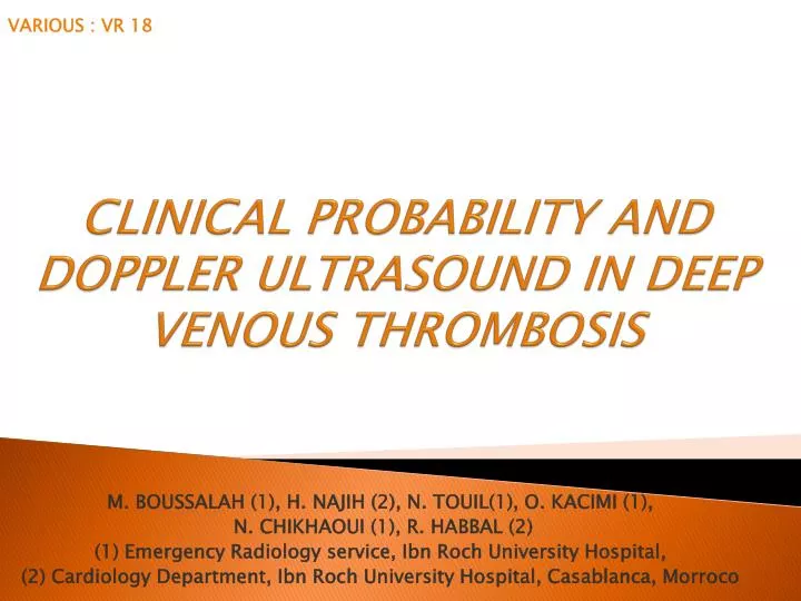
CLINICAL PROBABILITY AND DOPPLER ULTRASOUND IN DEEP VENOUS THROMBOSIS. M. BOUSSALAH (1), H. NAJIH (2), N. TOUIL (1), O. KACIMI (1 ), N. CHIKHAOUI (1), R. HABBAL (2 ) (1) Emergency Radiology service, Ibn Roch University Hospital ,

E N D
CLINICALPROBABILITY AND DOPPLERULTRASOUND IN DEEPVENOUSTHROMBOSIS M. BOUSSALAH (1), H. NAJIH (2), N. TOUIL(1), O. KACIMI (1), N. CHIKHAOUI (1), R. HABBAL (2) (1) Emergency Radiology service, Ibn Roch UniversityHospital, (2) CardiologyDepartment, Ibn Roch UniversityHospital, Casablanca, Morroco VARIOUS : VR 18
INTRODUCTION : • Clinical assessment is a fundamental step in the diagnostic workup of patients with suspected deep venous thrombosis (DVT). • Although the diagnostic yield of individual symptoms, signs, and common laboratory tests are limited, the combination of these variables can be used to express a clinical probability of DVT. • It is important to asses it because treatment must be undertaken rapidly to avoid potentially fatal pulmonary embolism [1]. • Based on history and physical examination for predicting the probability of deep venous thrombosis, clinical scores were developed and improved the efficiency of early detection [2, 3]. VARIOUS : VR 18
POPULATION AND METHODS : • Prospective study during 10 March 2010 to 10 April 2011, • Including 124 patients admitted in the emergency department with clinical supposition of lower extremity DVT, • The Wells score (Table. 1) was calculated : • high probability of DVT if the score is >3 points; • moderate probability if the score is 1 or 2 points; • low probability if <0 points, VARIOUS : VR 18
POPULATION AND METHODS : • An ultrasound examination was performed in all cases, using : 3-5 MHz convex transductor (for iliac veins) and 7.5 MHz linear transductor (from femoral level to distal). • Finding for DVT diagnosis were considered to be the following : • identification of a thrombus (with different degrees of echogenicity depending upon the moment of formation, • lack of compressibility of the vein, • distension of the recent thrombosed vein. VARIOUS : VR 18
POPULATION AND METHODS : • Doppler finding included : • decreased flow augmentation proximal to the occlusive thrombus, • continuous flow distal to the affectedvein, • absence or diminution of Valsalva response. • By color flow in non-occlusive thrombus, the blood flow around the thrombus was analyzed. In vein occlusion, the collateral tiny channels adjacent to the vein wall was searched. • The statistical method use SPSS 16.0 vr F; P < 0.05 was considered significant statistically. VARIOUS : VR 18
POPULATION AND METHODS : Table. 1 : Wells score [2]. VARIOUS : VR 18
RESULTS : EPIDEMIOLOGY • Mean age : 49 years old • Deviation standard : 17 • Sex distribution : 42 men and 82 women (fig. 1), • sex ratio male/female: 0.51 Fig. 1 : Distribution of the patients basedupon sex. VARIOUS : VR 18
RESULTS : EPIDEMIOLOGY Fig. 2 : The distribution of the patients basedupon âge decade VARIOUS : VR 18
RESULTS : CLINICAL PRESENTATION • The patients present clinical signs of DVT : • Unilateral pitting oedema found in 54.8% • Calf pain and tenderness : 62% • Phlegmasia alba dolens : 1% • Phlegmasiacaeruleadolens : 1% . • The DVT was confirmed in : 64.51% (80 patients) • Right lower limb : 28.2% , • Left lower limb : 27.41%, • Both lower limb : 7.25%. • Lower limb DVT localization : • Proximal : 30.64%, • Distal : 9.6%, • Total : 24.2%. VARIOUS : VR 18
RESULTS : WELLS SCORE • Based upon Wells score results the patients were divided into 3 groups (Fig. 3) : • First group – Low Probability: 26 patients, • Second group – Moderate probability : 42 patients, • Third group – High probability : 56 patients. Fig. 3: Distribution of the patients based upon Wells Score. VARIOUS : VR 18
RESULTS : DOPPLER US • Doppler ultrasound examination was performed in all cases (Fig. 4) : • Diagnosis of DVT : 80 patients, • Normal : 44 patients. Fig. 4: Distribution of doppler ultrasound finding in our study. VARIOUS : VR 18
RESULTS : DOPPLER US VERSUS WELLS SCORE Fig. 5 : Clinical probability, based on Wells Score, confronted to dopller US finding in our study. VARIOUS : VR 18
RESULTS : DOPPLER US VERSUS WELLS SCORE • Significant difference regarding the DVT diagnosis was found : • Third - second group : P = 0,0005 • Second - first group : P = 0,0001 VARIOUS : VR 18
DISCUSSION : DVT • DVTis the formation of a blood clot within a deep vein, and occurs about 1/1000 per year [4]. • It may occur in upper limb veins including the subclavian vein, visceral veins or the vena cava. More commonly, it occurs in the deep veins of the lower leg or the proximal veins of the ilio-femoral segment. • It is associated with a risk of pulmonary embolism (PE), making it potentially dangerous and an immediate diagnosis essential. • The diagnosis or reliable exclusion of DVT is a daily task for clinicians. Yet, the approach used is often not standardized and sometimes lacks effectiveness and accuracy due to the wide range of possible tests, the heterogeneity of the various tests available, and the different experiences of physicians requesting or performing these examinations. VARIOUS : VR 18
DIAGNOSISPROCEDURES • Clinical examination : • Its clinical symptoms can include leg swelling, pain, and erythema, which result from outflow obstruction and venous wall inflammation. • It, however, varies from minimal symptoms to massive features, and it is commonly believed that this variability is unreliable in predicting the diagnosis of DVT in patients with leg symptoms, leading to a large number of negative studies with venous duplex scanning [5]. • Thus, physical examination alone is not sufficient and should be followed by more sensitive and specific testing. VARIOUS : VR 18
DIAGNOSISPROCEDURES • Pretest probability scores : • Recently, clinical assessment in the form of scoring systems, while unable to exclude or confirm the presence of DVT, has gained favor as a method of stratifying pretest probability [6, 7]. • The best-known clinical scoring system was developed by Wells et al. and has been validated by other authors [8]. • This scoring system assigns a numeric score to patients based on certain set of validation DVT risk factors (Table. 1). • Patients are determined to be at low, moderate or high risk. • The incidence of DVT in these groups ranges from 3% to 13%, 17% to 38%, and 60% to 75%, respectively, also, the difference in prevalence of DVT in the three categories was statistically significant [5,9]. VARIOUS : VR 18
DIAGNOSISPROCEDURES • Pretest probability scores : • In 2003, Wells et al. published a lightly different score wish again included previously documented DVT and divided patients into only two groups, one in wish DVT was likely (score > 2) and one in wish it was unlikely (score < 2) [4]. • Our studyagreed with the literature on pretest probability of the DVT, by demonstrating the the Wells score was useful in estimating the probability of DVT. VARIOUS : VR 18
DIAGNOSISPROCEDURES • D-dimer test : • D-dimer is a degradation product of cross-linked fibrin, and elevated levels indicate fibrinolysis. • Increased D-dimer are marker of thromboembolic events, however, its specificity is low. • Different D-dimer tests are available and all assays vary in performance characteristics [4]. • The importance of D-dimer testing lies in the negative predictive value. Normal values effectively rule out PE. The negative predictive value for suspected DVT is lower than for PE, but in connection with pretest probability scores extensively evaluated [4]. VARIOUS : VR 18
DIAGNOSISPROCEDURES • Ultrasound examination : • When the measurement of the D-dimers cannot be performed, Doppler US represents the test of choice for the confirmation of DVT diagnosis [10]. • Several studies have reported a sensitivity of 95% and specificity of 98% of this method for the DVTdiagnosis, especially for proximal thrombosis [11]. • The overall sensitivity of ultrasound is considered to be between 86-98% [10]. • Ultrasound is less sensitive in patients with distal DVT; a negative ultrasound does not rule out the diagnosis of DVT at this level. VARIOUS : VR 18
DIAGNOSISPROCEDURES • Ultrasound examination : • In the absence of DVT, the veins collapse with a complete apposition of the vein walls during gentle compression. The loss of this property is the principal criterion for the diagnosis of DVT. • According to several studies, patients with a high probability of DVT have over a 75% prevalence of DVT confirmed by tests [9]. • We obtained similar results in our study in cases with a high probability : 64.5% patients with high probability had a confirmed DVT. VARIOUS : VR 18
DIAGNOSISPROCEDURES • Others procedures : • The venography is still the gold standard. However, radiation, contrast media, and the painful injection in pedal veins are limiting factors [4]. • Spiral CT venography has been shown to have a sensitivity and specificity similar to that of conventional venography. However, costs and high radiation are limiting factors for the routine use of venous imaging [4]. • Recent studies show a high sensitivity and specificity for MR venography, but its high costs and limited availability exclude it from routine use [4, 12]. VARIOUS : VR 18
Diagnosisstrategies • As an initial step in the diagnosis of DVT, the pretest probability score is recommended using the defined protocols. • The patients should undergo either ultrasound or D-dimer testing (using a D-dimer test with a high sensitivity) [4]. • Subsequent steps should depend on the outcome of the pretest probability evaluation and the results. VARIOUS : VR 18
Diagnosisstrategies Fig. 6 : Proposed strategy for patients with suspected deep vein thrombosis (DVT). [5] Non-high clinical score: low or moderate clinical score; PCP, pretest clinical probability. VARIOUS : VR 18
REFERENCES : • Lindblad B, Sternby NH, Bergqvist D. Incidence of venousthromboembolismverifiedby necropsy over 30 years.BMJ 1991; 302: 709-11. • Wells PS, Hirsh J, Anderson DR, LensingAW, Foster G, KearonC, et al.Accuracy of clinicalassessment of deep-veinthrombosis.Lancet 1995; 345:1326-30 • Constans J, Boutinet C, Salmi L.R, SabyJ.C, NelzyM.L, Baudouin P, et al. Comparison of four clinicalprediction scores for the diagnosis of lowerlimbdeepvenousthrombosis in outpatients.AmJ Med 2003; 115: 436-40 • Beyer J, Schellong S, Deepveinthrombosis: Currentdiagnosisstrategy. European Journal of InternalMedicine 2005; 16 : 238-246. • Ymaki T, Nozaki M, Sakurai H, Takeuchi M, Soejima K, Kono T, Prospective Evaluation of a Screening Protocol to Exclude Deep Vein Thrombosis on the Basis of a Combination of Quantitative D-Dimer Testing and Pretest Clinical Probability Score. J Am CollSurg; 2005; 201:1: 701-709. • Zhu L, Guo X, Wang J, Guo Y, Wang C, Ma H, Guo Y, Comparison of four clinical scores for the predicting lower limb deep venous thrombosis in Chinese patients Journal of Nanjing MedicalUniversity, 2008, 22(4): 230-233 VARIOUS : VR 18
REFERENCES : • Delluc A, Le Pape F, Le Bras A, Gagne P, Taton G, Jaffrelot M, Le Duff N, Bressollette L, Le Gal G, Validation d’un score de prédiction clinique de la thrombose veineuse profonde des membres inférieurs spécifique à la médecine générale. Rev Med Interne (2012), doi:10.1016/j.revmed.2011.12.004 • Ambid-Lacombe C, CambouJP, Bataille V, Baudoin D, Vassal-Hebrard B, Boccalon H, Bura Rivière A, Excellentes performances du score de Wells et du score de Wells modifié dans le diagnostic de thrombose veineuse profonde proximale ou distale chez des patients hospitalisés ou ambulatoires au CHU de Toulouse : étude TVP-PREDICT. Journal des Maladies Vasculaires 2009; 34 : 211—217. • Tracy JA, Edlow JA, Ultrasound diagnosis of deep venous thrombosis.EmergMed Clin N Am 22 (2004) 775–796. • Hotoleanu C, Fodor D, Suciu O, Correlations between clinical probability and Doppler ultrasound results in the assessment of deep venous thrombosis, Medical Ultrasonography; 2010,;12; 1: 17-21. • Segal JB, Eng J, TamarizLJ, Bass EB. Review of the evidence on diagnosis of deep venous thrombosis and pulmonary embolism. Ann Fam Med 2007; 5: 63-7. • Evans AJ, SostmanHD, KnelsonMH, Spritzer CE, Newman GE, Paine SS, et al. 1992 ARRSExecutive Council Award. Detectionof deepvenousthrombosis: prospective comparison of MR imagingwithcontrastvenography. Am J Roentgenol 1993;161(1):131–9 (Jul). VARIOUS : VR 18
CONCLUSION : • The clinical diagnosis of deep venous thrombosis, which has been viewed as a clinical challenge for many years, has been improved through the use of the standardized assessment of the clinical probability of deep venous thrombosis before performing a diagnostic test. • Thus, the combination of pretest probability with non-invasive diagnostic test results simplifies and improves the diagnostic process in patients with suspected low extremity deep-vein thrombosis, and will decrease costs. • Us showed in our study, asignificant correlation between the clinical probability of deep venous thrombosis and the proportion of cases confirmed by doppler ultrasound. VARIOUS : VR 18
ABSTRACT : • Objectives :Deep Venous Thrombosis (DVT) is associated with a significant morbidity and mortality. Its diagnosis is based on clinical examination and complementary investigation, especially Doppler ultrasound. We aim to establish the correlation between the clinical probabilities of DVT based on Wells score and the results of Doppler ultrasound exam. • Materials and methods : We describe findings in a 124 patients with clinical supposition of DVT divided into 3 groups based upon the probability of DVT (Wells score): low, moderate and high, respectively. All the patients were examined by Doppler ultrasonography. VARIOUS : VR 18
ABSTRACT : • Results : The DVT was confirmed by Doppler ultrasonography in more than half of the cases; the highest percent of confirmed cases were in the patients with a high probability of DVT (64, 5%) whereas the lowest percent was associated with the low clinical probability (20%). There was a significant difference regarding the DVT diagnosis between third and the second group (p= 0, 0005), also between the second and the first group (p= 0, 0001). • Conclusion :In the diagnosis of deep venous thrombosis, an exhaustive doppler ultrasound exam is necessary when the clinical probability is high, based on the Wells score. A significant correlation between the clinical probability of DVT and the proportion of cases confirmed by ultrasound, was demonstrated in this study. VARIOUS : VR 18