Download
1 / 28
280 likes | 301 Vues
Delve into diagnosing and managing pulmonary embolism with tips, clinical scores, investigations, and implications from literature and modern practices.

E N D
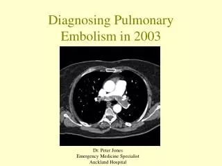
Diagnosing Pulmonary Embolism in 2003 Dr. Peter Jones Emergency Medicine Specialist Auckland Hospital
Case 143f • 5 days post LUSCS • Sudden onset pleuritic R chest pain • HR 104 • WCC 13, D-Dimer <500 -> 500-4000 3/7 later • CxR blunting R CP angle • CTPA negative • Diagnosis: pleurisy • Subsequently well
Case 2 19f smoker • Dry cough and pleuritic central chest pain <1/7 • RR 20 • ABG • pO2 10.3kPa (77mmHg) • A-a 3.7kPa (24mmHg – expected 9mmHg) • D-Dimer <500, CxR normal, ECG normal • CTPA negative, no positive (maybe) • Anticoagulated 5 days • V/Q ‘low probability’ day 4 • Diagnosis: Chest pain ?cause
Case 329f • 1/12 ago “blackout” • 3/7 SOB and central chest pain (not pleuritic) • 2/7 ago right leg cramp • Occasional crepitation right base • ECG RBBB, CxR normal • ABG • pO2 10kPa (75mmHg) • A-a 3.3kPa (21mmHg – expected 11mmHg) • D-Dimer <500 • Medical referral • Tender R costochondral junction,”flu” 1/12 ago • Diagnosis: Costochondritis
Background PE • Incidence: 23/100000/yr • Mortality 30% at 1 yr untreated, 2-8% treated • 50% undiagnosed pre mortem • Wide differential diagnosis • Poor clinical signs • Myriad of diagnostic strategies • No consensus
Massive PEBTS 2003 • Highly Likely If • Collapse / hypotension and • Unexplained hypoxia and • Engorged neck veins and • RV Gallop • Bedside echocardiogram or CTPA <1hr
Non-massive PEThe Literature • Using a pre-investigation clinical score is useful • Defines a group of low risk patients that it is safe to use D-Dimer as a ‘rule out’ test • Use Bayes’ theorem to calculate post test probability • The Se and Sp of each test needs to be known, these used to determine likelihood of PE given + or - test • Post test probability of <2% accepted as sufficient to stop investigating for PE and look for other cause • Post test probability >80% treat
Bayes’ TheoremEssay Towards Solving a Problem in the Doctrine of ChancesPhilosophical Transactions of the Royal Society of London (1764). • Bayes theorem • Take pre test probability • Calculate odds • Multiply by test Likelihood Ratio to get post test odds • Calculate post test probability Thomas Bayes 1702-1761
PE Clinical Scores 1. Empiric (Best guess - Auckland) 2. Derived from logistic regression analysis of known risk factors, clinical and investigation findings - complex derivation • Wells et al. (Canada) • Wicki et al. (Switzerland) • Kline et al. (USA)
Wells et al. Clinical Model SimplifiedThrombosis Haemostasis 2000; 83:416-20 976 patients, prevalence PE 17% 3 Clinical Sn/Sx DVT Minimum leg swelling + pain on palpation deep veins 3 Alternative diagnosis less likely than PE 1.5 HR >100 1.5 Immobilisation or surgery previous 4 weeks 1.5 Previous VTE 1 Haemoptysis 1 Malignancy Treated in last 6/12 or palliative
Score %Pts %PE <2 Low 40 3.6 2-6 Mod 53 20.5 >6 High 7 66.7 <=4 Unlikely 71 7.8 >4 Likely 29 40.7 Wells et al. >2500 patients several studies
Wicki et al. Arch. Int. Med. 2001:161, 92-98986 ED patients prevalence 27% Score Risk %Pts %PE <4 Low 49 10 4-8 Int. 44 38 >8 High 6 81
Category %Pts %PE Safe 79 13.3 Unsafe 21 42 HR/SBP >1 or Age > 50 sO2 < 95% (normal lungs) Unilateral leg swelling Recent surgery Haemoptysis Kline et al Ann Emerg. Med. 2002:39:2, 144-152934 ED patients, prevalence of PE 19% If yes to any of the above, asked in this order, then ‘unsafe’ to test with D-Dimer
PE InvestigationsD-Dimer Assays use an antibody directed against the D-dimer peptide, which is a product of fibrin breakdown • Currently using latex agglutination test • Sufficiently inaccurate so that post test probability 9% in low risk patients, 60% in high risk (empiric score) • Negative LR 0.38, Positive LR 3.5 • Rapid ELISA test now available (Vidas) • Negative LR 0.07, Positive LR 1.6 • Post test probability <2% in low risk group, 23% in high risk group
PE Investigations CTPA • Multitude of papers • Modern multi-slice scanners accurate • Probably as good as pulmonary angiography • Sn 95%, Sp95% • -ve LR 0.06, +ve LR 19 • Can reveal other diagnosis • Some scans will be equivocal • Relative contraindications • Renal impairment • Young women • Absolute contraindication • Contrast Allergy
NO LEG SIGNS 3 studies Approx 2000 patients NLR 0.39-0.73 LEG SIGNS 5 Studies >2000 patients NLR 0.02-0.16 PE InvestigationsCompression Ultrasound
Investigations • Pulmonary Angiography • ?Consigned to history • VQ scan • May have a place if CTPA contraindicated • NRL 0.1 • only 14% have normal/near normal scans • 9% of these had PE in PIOPED
Algorithms • Plethora of published algorithms with various strategies • Aim to rule out PE with post test probability <2% • Recently published (2003) • British Thoracic Society • Emergency Medicine
Currently at Auckland Applying Bayes’ Theorem to the presented cases, using our current tests
RMO Handbook 4% 6%
Ignoring the Pretest Probability • Parallel stream in the literature • Assumptions • “A negative Vidas D-Dimer rules out PE” • “A negative CTPA rules out PE” • Problems • Vidas D-Dimer will miss PE • Sensitivity 90-100%, NLR 0.05-0.22 • 50-85% of patients require CTPA, 5-23% have PE • CTPA will miss PE / be equivocal • Sensitivity 95%, specificity 95%, NLR 0.06
Proposed Algorithm Well’s Score 4 or less Unlikely PE Well’s Score >4 Likely PE CTPA / VQ Vidas D-Dimer + - Well’s likely Well’s Unlikely Compression US + - - + 2.2% CTPA 3.6% VQ Other cause Post test prob > 75% Treat
Algorithm Implications Regardless of chosen algorithm, imaging for PE will increase markedly (diagnosis of PE will also increase)
Thomas Bayes 1736 “..why, therefore, if I am half blind, must I take for my guide one that cannot see at all?”
Selected References • Clinical Score PE • Wells PS et Al. Thromb. Haemost. 2000: 83;416-20 • Wicki et Al Arch. Int. Med. 2001:161, 92-98 • Kline et al Ann Emerg. Med. 2002:39:2, 144-152 DVT • Wells PS et al. Thromb. Haemost. 1999 81 493-7 • D-Dimer • Kline paper above (good summary of the issues) • Sijens PE et al. Thromb. Haemost. 2000: 84; 156-9 • CTPA vs Pulmonary Angio • Wells PS et al. Clin Chest Med 24 2003 13-28 Chapter ‘Diagnosis of PE: When is imaging needed?’ • Kline et al. Ann Emerg Med 35 (2) 2000; 168-80 • Compression Ultrasound • Turkstra et al. Ann Int med 1997 126(10) 775-781 • Heijboer H et al. NEJM 1993 329 (19) 1365-1369 • Perrier A et Al Lancet 1999 353 190-195 • Strategies PE • Perrier A et al Lancet 1999 353 190-195 • Musset et al. Lancet 2002 360 1914-1920 • Chagnon et al. Am J Med 2002 113: 269-275 • BTS Standards of care committee Thorax 2003 58 470-83 • British Thoracic Society Guidelines • Mountain D. emergency medicine 2003 15 (3); 250-62 • Fedullo PF et al. NEJM 2003 349 (13); 1247-56 • Kline JA and Wells PS: Ann Emerg Med August 2003 42:2 266-276 DVT • Wells et al NEJM 2003 349 (13); 1227-35 (and many others)


