Download
1 / 82
940 likes | 1.53k Vues
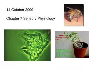
Sensory Physiology. Nervous System - Senses. General Senses receptors that are widely distributed throughout the body skin, various organs and joints Special Senses specialized receptors confied to structures in the head eyes and ears. Senses. Sensory Receptors

E N D
Nervous System - Senses • General Senses • receptors that are widely distributed throughout the body • skin, various organs and joints • Special Senses • specialized receptors confied to structures in the head • eyes and ears
Senses • Sensory Receptors • specialized cells or multicellular structures that collect information from the environment • stimulate neurons to send impulses along sensory fibers to the brain • Sensation • a feeling that occurs when brain becomes aware of sensory impulse • Perception • a person’s view of the stimulus; the way the brain interprets the information
Sensory Receptor Types Figure 10-1: Sensory receptors
Receptor Types • Chemoreceptors • respond to changes in chemical concentrations • Pain receptors (Nociceptors) • respond to tissue damage • Thermoreceptors • respond to changes in temperature • Mechanoreceptors • respond to mechanical forces • Photoreceptors • respond to light
Sensory Adaptation • ability to ignore unimportant stimuli • involves a decreased response to a particular stimulus from the receptors (peripheral adaptations) or along the CNS pathways leading to the cerebral cortex (central adaptation) • sensory impulses become less frequent and may cease • stronger stimulus is required to trigger impulses
General Senses • senses associated with skin, muscles, joints, and viscera • three groups • exteroceptive senses – senses associated with body surface; touch, pressure, temperature, pain • visceroceptive senses – senses associated with changes in viscera; blood pressure stretching blood vessels, ingesting a meal • proprioceptive senses – senses associated with changes in muscles and tendons
Touch and Pressure Senses • Free nerve endings • common in epithelial tissues • simplest receptors • sense itching • Ex ( nociciptor) • Meissner’s corpuscles • abundant in hairless portions of skin; lips • detect fine touch; distinguish between two points on the skin • Pacinian corpuscles • common in deeper subcutaneous tissues, tendons, and ligaments • detect heavy pressure and vibrations
Touch (pressure) Figure 10-11: Touch-pressure receptors
Temperature Senses • Warm receptors • sensitive to temperatures above 25oC (77o F) • unresponsive to temperature above 45oC (113oF) • Cold receptors • sensitive to temperature between 10oC (50oF) and 20oC (68oF) • Pain receptors (nociceptor) • respond to temperatures below 10oC • respond to temperatures above 45oC
Special Senses • sensory receptors are within large, complex sensory organs in the head • smell in olfactory organs • taste in taste buds • hearing and equilibrium in ears • sight in eyes
Sense of Smell • Olfactory Receptors • chemoreceptors • respond to chemicals dissolved in liquids • Olfactory Organs • contain olfactory receptors and supporting epithelial cells • cover parts of nasal cavity, superior nasal conchae, and a portion of the nasal septum
Olfactor: Sense of Smell Figure 10-14a, b: ANATOMY SUMMARY: Olfaction
Olfactor: Sense of Smell Figure 10-14c: ANATOMY SUMMARY: Olfaction
Olfactory Nerve Pathways • Once olfactory receptors are stimulated, nerve impulses travel through • olfactory nerves olfactory bulbs olfactory tracts limbic system (for emotions) and olfactory cortex (for interpretation)
Olfactory Stimulation • olfactory organs located high in the nasal cavity above the usual pathway of inhaled air • olfactory receptors undergo sensory adaptation rapidly • sense of smell drops by 50% within a second after stimulation • Olfactory Code • hypothesis • odor that is stimulated by a distinct set of receptor cells and its associated receptor proteins
Sense of Taste • Taste Buds • organs of taste • located on papillae of tongue, roof of mouth, linings of cheeks and walls of pharynx • Taste Receptors • chemoreceptors • taste cells – modified epithelial cells that function as receptors • taste hairs –microvilli that protrude from taste cells; sensitive parts of taste cells
Taste Sensations • Four Primary Taste Sensations • sweet – stimulated by carbohydrates • sour – stimulated by acids • salty – stimulated by salts • bitter – stimulated by many organic compounds Spicy foods activate pain receptors
Taste Nerve Pathways • Sensory impulses from taste receptors travel along • cranial nerves to • medulla oblongata to • thalamus to • gustatory cortex (for interpretation)
Taste: Chemoreceptors • Salt --- Na passive • Acids– H block k channels • Sweet – Gs anhydrate cyclase – protein kinase(A) – Phosphorylation of k channels so it will close • Bitter – Gb – phospholipasec – release of ca from ER • Depolarization –activate neurotransmitter vesicle to diffues to membrane and release neurotransmitter
The Ear: Hearing and Equilibrium • The ear – receptor organ for hearing and equilibrium • Composed of three main regions • Outer ear – functions in hearing • Middle ear – functions in hearing • Inner ear – functions in both hearing and equilibrium
The Outer (External) Ear • Composed of: • The auricle (pinna) • Helps direct sounds • External acoustic meatus • Lined with skin • Contains hairs, sebaceous glands, and ceruminous glands • Tympanic membrane • Forms the boundary between the external and middle ear
The Outer (External) Ear Figure 16.17a
The Middle Ear • The tympanic cavity • A small, air-filled space • Located within the petrous portion of the temporal bone • Medial wall is penetrated by: • Oval window • Round window • Pharyngotympanic tube (auditory or eustachian tube) • Links the middle ear and pharynx
Structures of the Middle Ear Figure 16.17b
The Middle Ear • Ear ossicles – smallest bones in the body • Malleus – attaches to the eardrum • Incus – between the malleus and stapes • Stapes – vibrates against the oval window Figure 16.19
The Inner (Internal) Ear • Inner ear – also called the labyrinth • Lies within the petrous portion of the temporal bone • Bony labyrinth – a cavity consisting of three parts • Semicircular canals • Vestibule • Cochlea
The Inner (Internal) Ear • Membranous labyrinth • Series of membrane-walled sacs and ducts • Fit within the bony labyrinth • Consists of three main parts • Semicircular ducts • Utricle and saccule • Cochlear duct
The Inner (Internal) Ear • Membranous labyrinth (continued) • Filled with a clear fluid – endolymph • Confined to the membranous labyrinth • Bony labyrinth is filled with perilymph • Continuous with cerebrospinal fluid
The Membranous Labyrinth Figure 16.20
The Inner (Internal) Ear Figure 16.17b
Cochlea • Cochlear duct • portion of membranous labyrinth in cochlea • Vestibular membrane • separates cochlear duct from scala vestibuli • Basilar membrane • separates cochlear duct from scala tympani
The Cochlea Figure 16.23a–c
Organ of Corti • group of hearing receptor cells (hair cells) • on upper surface of basilar membrane • different frequencies of vibration move different parts of basilar membrane • particular sound frequencies cause hairs of receptor cells to bend • nerve impulse generated
Hearing: Mechanoreceptors Figure 10-19: Sound transmission through the ear
Hearing: Hair Cell Transduction Figure 10-20: The cochlea
Hearing: Hair Cell Transduction Figure 10-21: Signal transduction in hair cells
Equilibrium • Dynamic Equilibrium • semicircular canals • sense rotation and movement of head and body • Static Equilibrium • vestibule • sense position of head when body is not moving
Vestibule • Utricle • communicates with saccule and membranous portion of semicircular canals • Saccule • communicates with cochlear duct • Mucula • hair cells of utricle and saccule
Macula • responds to changes in head position • bending of hairs results in generation of nerve impulse