Download
1 / 69
750 likes | 1.21k Vues
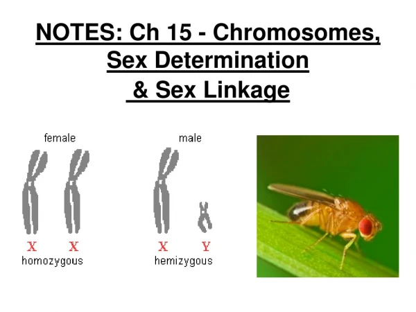
The Sex Chromosomes and Their Abnormalities. The Chromosomal Basis of Sex Determination. The fundamental basis of the XX/XY system of sex determination: Males with Klinefelter syndrome: karyotype 47,XXY, whereas most Turner syndrome females have only 45 chromosomes with a single X chromosome.

E N D
The Chromosomal Basis of Sex Determination The fundamental basis of the XX/XY system of sex determination: • Males with Klinefelter syndrome: karyotype 47,XXY, whereas most Turner syndrome females have only 45 chromosomes with a single X chromosome. • The the Y chromosome is crucial in normal male development. • Furthermore, the effectof varying number of X chromosomes is moderate in either males or females.
In addition to sex chromosomes, a number of genes located on both the sex chromosomes and the autosomes are involved in sex determination and subsequent sexual differentiation. • In most instances, the role of these genes has come to light as a result of patients with abnormalities in sexual development, whether cytogenetic, mendelian, or sporadic.
The structure of the Y chromosome and its role in sexual development have been determined at both the molecular and genomic levels. • In male meiosis, the X and Y chromosomes normally pair by segments and undergo recombination at the pseudoautosomal region of the X and Y chromosomes. • The Y chromosome is relatively gene poor and contains only about 50 genes. The functions of a high proportion of these genes are related to gonadal and genital development.
The Y chromosome in sex determination and in disorders of sexual differentiation. Individual genes and regions implicated in sex determination, sex reversal, and defects of spermatogenesis are indicated.
Embryology of the Reproductive System • By sixth week of development in both sexes, the primordial germ cells have migrated from their earlier extraembryonic location to the gonadal ridges, where they are surrounded by the sex cords to form a pair of primitive gonads. The developing gonad is bipotential and is often referred to as indifferent. • The development into an ovary or a testis is determined by the coordinated action of a sequence of genes that leads normally to ovarian development when no Y chromosome is present or to testicular development when a Y is present. • The ovarian pathway is followed unless a Y-linked gene, designated testis-determining factor (TDF), acts as a switch, diverting development into the male pathway.
Scheme of developmental events in sex determination and differentiation of the male and female gonads. Involvement of individual genes in key developmental steps or in genetic disorders is indicated in blue boxes.
The Testis-Determining Gene, SRY • Whereas the X and Y chromosomes normally exchange in meiosis I within the Xp/Yp PAR, in rare instances, genetic recombination occurs outside of the PAR, leading to two rare but highly informative abnormalities: XX males and XY females. • Each of these sex-reversal disorders occurs with an incidence of approximately 1 in 20,000 births.
XX males are phenotypic males with a 46,XX karyotype who usually possess some Y chromosomal sequences translocated to the short arm of the X. • Similarly, a proportion of phenotypic females with a 46,XY karyotype have lost the testis-determining region of the Y chromosome. • The SRY gene (sex-determining region on the Y) lies near the pseudoautosomal boundary on the Y chromosome, is present in many 46,XX males, and is deleted or mutated in a proportion of female 46,XY patients, thus strongly implicating SRY in male sex determination.
SRY is expressed only briefly early in development in cells of the germinal ridge just before differentiation of the testis. • SRY encodes a DNA-binding protein that is likely to be a transcription factor, although the specific genes that it regulates are unknown. SRY is equivalent to the TDF gene on the Y chromosome. • However, the presence or absence of SRY does not explain all cases of abnormal sex determination. SRY is not present in about 10% of unambiguous XX males and in most cases of XX true hermaphrodites or XX males with ambiguous genitalia. • Further, mutations in the SRY gene account for only about 15% of 46,XY females. Thus, other genes are implicated in the sex-determination pathway.
Figure 6-12 Etiological factors of XX male or XY female phenotypes by aberrant exchange between X- and Y-linked sequences.
Y-Linked Genes in Spermatogenesis • Interstitial deletions in Yq have been associated with at least 10% of cases of nonobstructive azoospermia and with approximately 6% of cases of severe oligospermia. • These findings suggest that one or more genes, termed azoospermia factors (AZF), are located on the Y chromosome, and three nonoverlapping regions on Yq (AZFa, AZFb, and AZFc) have been defined.
Molecular analysis of these deletions has led to identification of a series of genes that may be important in spermatogenesis. E.g., the AZFc deletion region contains several families of genes expressed in the testis, including the DAZ genes (deleted in azoospermia) that encode RNA-binding proteins expressed only in the premeiotic germ cells of the testis. • De novo deletions of AZFc arise in about 1 in 4000 males and are mediated by recombination b/w long repeated sequences.
AZFa and AZFb deletions, although less common, also involve recombination. • Approximately 2% of otherwise healthy males are infertile because of severe defects in sperm production, and it appears likely that de novo deletions or mutations account for at least a proportion of these.
Thus, men with idiopathic infertility should be karyotyped, and Y chromosome molecular testing and genetic counseling may be appropriate before the initiation of assisted reproduction for such couples. • Not all cases of male infertility are due to chromosomal deletions. For example, a de novo point mutation has been described in one Y-linked gene, USP9Y, the function of which is unknown but which must be required for normal spermatogenesis.
The X ChromosomeChromosome Inactivation • The relative tolerance of the human karyotype for X chromosome abnormalities can be explained in terms of X chromosome inactivation, the process by which most genes on one of the two X chromosomes in females are silenced epigenetically and fail to produce any product. • The theory of X inactivation is that in somatic cells in normal females, one X chromosome is inactivated early in development, thus equalizing the expression of X-linked genes in the two sexes.
Figure 6-15 Profile of gene expression of the X chromosome. Each symbol indicates X inactivation status of an X-linked gene. Location of each symbol indicates its approximate map position on the X chromosome. Genes not expressed from the inactive X (subject to inactivation) are on the left. Genes expressed from the inactive X (escape from inactivation) are on the right; genes represented in light blue are those that escape inactivation in only a subset of females tested. The location of the XIST gene and the X inactivation center (XIC) are indicated in Xq13.2.
Cytogenetic Abnormalities of the Sex Chromosomes • As a group, disorders of the sex chromosomes tend to occur as isolated events without apparent predisposing factors, except for an effect of late maternal age in the cases that originate from errors of maternal meiosis I. • clinical indications that raise the possibility of a sex chromosome abnormality and thus the need for cytogenetic or molecular studies include delay in onset of puberty, primary or secondary amenorrhea, infertility, and ambiguous genitalia.
X and Y chromosome aneuploidy is relatively common, and sex chromosome abnormalities are among the most common of all human genetic disorders, with an overall incidence of about 1 in 400 to 500 births. • The phenotypes associated with these chromosomal defects are, in general, less severe than those associated with comparable autosomal disorders because X chromosome inactivation, as well as the low gene contentof the Y, minimizes the clinical consequences of sex chromosome imbalance.
By far the most common sex chromosome defects in live-born infants and in fetuses are the trisomic types (XXY, XXX, and XYY), but all three are rare in spontaneous abortions. • In contrast, monosomy for the X (Turner syndrome) is less frequent in live-born infants but is the most common chromosome anomaly reported in spontaneous abortions.
Structural abnormalities of the sex chromosomes are less common; the defect most frequently observed is an isochromosome of the long arm of the X, i(Xq), seen in complete or mosaic form in at least 15% of females with Turner syndrome. • Mosaicism is more common for sex chromosome abnormalities than for autosomal abnormalities, and in some patients it is associated with relatively mild expression of the associated phenotype.
Incidence of Sex Chromosome Abnormalities Overall incidence: 1/650 females
The four well-defined syndromes associated with sex chromosome aneuploidy are important causes of infertility or abnormal development, or both. • As a group, those with sex chromosome aneuploidy show reduced levels of psychological adaptation, educational achievement, occupational performance, and economic independence, and on average, they scored slightly lower on intelligence (IQ) tests.
However, each group shows high variability, making it impossible to generalize to specific cases. In fact, the overall impression is a high degree of normalcy, particularly in adulthood, which is remarkable among those with chromosomal anomalies. • Because almost all patients with sex chromosome abnormalities have only mild developmental abnormalities, a parental decision regarding potential termination of a pregnancy in which the fetus is found to have this type of defect can be a very difficult one.
Klinefelter syndrome • Klinefelter syndrome patients are tall and thin and have relatively long legs. They appear physically normal until puberty, when signs of hypogonadism become obvious. • Puberty occurs at a normal age, but the testes remain small, and secondary sexual characteristics remain underdeveloped. • Gynecomastia is a feature of some patients; because of this, the risk of breast cancer is 20 to 50 times that of 46,XY males.
Klinefelter patients are almost always infertile because of the failure of germ cell development, and patients are often identified clinically for the first time because of infertility. • Klinefelter syndrome is relatively common among infertile males (about 3%) or males with oligospermia or azoospermia (5% to 10%). • In adulthood, persistent androgen deficiency may result in decreased muscle tone, a loss of libido, and decreased bone mineral density.
The incidence is at least 1 in 1000 male live births (1 in 2000 total births). Because of the relatively mild yet variable phenotype, many cases are presumed to go undetected. • About half the cases of Klinefelter syndrome result from errors in paternal meiosis I because of a failure of normal Xp/Yp recombination in the pseudoautosomal region. • Among cases of maternal origin, most result from errors in maternal meiosis I and the remainder from errors in meiosis II • or from a postzygotic mitotic error leading to mosaicism.
Maternal age is increased in the cases associated with maternal meiosis I errors. • About 15% of Klinefelter patients have mosaic karyotypes. As a group, such mosaic patients have variable phenotypes; some may have normal testicular development. • The most common mosaic karyotype is 46,XY/47,XXY, probably as a consequence of loss of one X chromosome in an XXY conceptus during an early postzygotic division.
There are several variants of Klinefelter syndrome, including 48,XXYY, 48,XXXY, and 49,XXXXY. As a rule, the additional X chromosomes (even though they are mostly inactive) cause a correspondingly more severe phenotype, with a greater degree of dysmorphism, more defective sexual development, and more severe mental impairment. • Although there is wide phenotypic variation, some consistent phenotypic differences have been identified between patients with Klinefelter syndrome and chromosomally normal males:
Verbal comprehension and ability are below those of normal males, and 47,XXY males score slightly lower on certain intelligence performance tests. • Patients with Klinefelter syndrome have a several-fold increased risk of learning difficulties, especially in reading, that may require educational intervention. • Klinefelter syndrome is over-represented among boys requiring special education. Many of the affected boys have relatively poor psychosocial adjustment, in part related to poor body image. Language difficulties may lead to shyness, unassertiveness, and immaturity
47,XYY Syndrome • Among all male live births, the incidence of the 47,XYY karyotype is about 1 in 1000. • The 47,XYY chromosome constitution is notassociated with an obviously abnormal phenotype • The origin of the error that leads to the XYY karyotype must be ????? • The less common XXYY and XXXYY variants, which share the features of the XYY and Klinefelter syndromes, probably also originate in the father as a result of sequential nondisjunction in meiosis I and meiosis II.
XYY males identified in newborn screening programs without ascertainment bias are tall and have an increased risk of educational or behavioral problems in comparison with chromosomally normal males. They have normal intelligence and are not dysmorphic. • Fertility is usually normal, and there appears to be no particularly increased risk that a 47,XYY male will have a chromosomally abnormal child. • About half of 47,XYY boys require educational intervention as a result of language delays and reading and spelling difficulties. Their IQ scores are about 10 to 15 points below average.
Parents whose child is found, prenatally or postnatally, to be XYY are often extremely concerned about the behavioral implications. • Attention deficits, hyperactivity, and impulsiveness have been well documented in XYY males. • reports in the 1960s and 1970s showed that the proportion of XYY males was elevated in prisons and mental hospitals, especially among the tallest inmates. This stereotypic impression is now known to be incorrect. • Nonetheless, inability to predict the outcome in individual cases makes identification of an XYY fetus one of the more difficult genetic counseling problems in prenatal diagnosis programs.
Trisomy X (47,XXX) • Trisomy X occurs with an incidence of 1 in 1000 female births. • Trisomy X females, although somewhat above average in stature, are not abnormal phenotypically. Some are first identified in infertility clinics, but probably most remain undiagnosed. • Follow-up studies have shown that XXX females develop pubertal changes at an appropriate age, and they are usually fertile although with a somewhat increased risk of chromosomally abnormal offspring.
There is a significant deficit in performance on IQ tests, and about 70% of the patients have some learning problems. • Severe psychopathological and antisocial behaviors appear to be rare; however, abnormal behavior is apparent, especially during the transition from adolescence to early adulthood
In 47,XXX cells, two of the X chromosomes are inactivated. Almost all cases result from errors in maternal meiosis, and of these, the majority are in meiosis I. • There is an effect of increased maternal age, restricted to those patients in whom the error was in maternal meiosis I. • The tetrasomy X syndrome (48,XXXX) is associated with more serious retardation in both physical and mental development. • The pentasomy X syndrome (49,XXXXX), despite the presence of four inactive X chromosomes, usually includes severe developmental retardation with multiple physical defects.
Turner Syndrome (45,X and Variants) • Unlike patients with other sex chromosome aneuploidies, females with Turner syndrome can often be identified at birth or before puberty by their distinctive phenotypic features. • Turner syndrome is much less common than other sex chromosome aneuploidies. The incidence of the Turner syndrome phenotype is approximately 1 in 4000 female live births.
The most frequent chromosome constitution in Turner syndrome is 45,X. • However, about 50% of cases have other karyotypes. • About one quarter of Turner syndrome cases involve mosaic karyotypes, in which only a proportion of cells are 45,X.
The most common karyotypes and their approximate relative prevalences are as follows:
The chromosome constitution is clinically significant. For example, patients with i(Xq) are similar to classic 45,X patients, whereas patients with a deletion of Xp have short stature and congenital malformations, and those with a deletion of Xq often have only gonadal dysfunction. • Typical abnormalities in Turner syndrome include short stature, gonadal dysgenesis (usually streak gonads reflecting a failure of ovarian maintenance), characteristic unusual faces, webbed neck, low posterior hairline, broad chest with widely spaced nipples, and elevated frequency of renal and cardiovascular anomalies. At birth, infants with this syndrome often have edema of the dorsum of the foot, a useful diagnostic sign.
Many patients have coarctation of the aorta, and Turner syndrome females are at particular risk for cardiovascular abnormalities. Lymphedema may be present in fetal life, causing cystic hygroma (visible by ultrasonography), which is the cause of the neck webbing seen postnatally. • Turner syndrome should be suspected in any newborn female with edema of the hands and feet or with hypoplastic left-sided heart or coarctation of the aorta. • The diagnosis should also be considered in the teenage years for girls with primary or secondary amenorrhea, especially if they are of short stature. Growth hormone therapy should be considered for all girls with Turner syndrome and can result in gains of 6 to 12 cm to the final height.
Intelligence in Turner syndrome females is usually considered to be normal, although approximately 10% of patients will show significant developmental delay requiring special education. • Even among those with normal intelligence, however, patients often display a deficiency in spatial perception, perceptual motor organization, or fine motor execution. • As a consequence, the nonverbal IQ score is significantly lower than the verbal IQ score, and many patients require educational intervention, especially in mathematics.
Turner syndrome females have an elevated risk of impaired social adjustment. A comparison of 45,X girls with a maternal X and those with a paternal X provided evidence of significantly worse social cognition skills in those with a maternally-derived X. • The high incidence of a 45,X karyotype in spontaneous abortions has already been mentioned. This single abnormality is present in an estimated 1% to 2% of all conceptuses; survival to term is a rare outcome, and more than 99% of such fetuses abort spontaneously.
The single X is maternal in origin in about 70% of cases; in other words, the chromosome error leading to loss of a sex chromosome is usually paternal. • Furthermore, it is not clear why the 45,X karyotype is usually lethal in utero but is apparently fully compatible with postnatal survival. • The "missing" genes responsible for the Turner syndrome phenotype must reside on both the X and Y chromosomes. It has been suggested that the responsible genes are among those that escape X chromosome inactivation, particularly on Xp, including those in the pseudoautosomal region.
Small ring X chromosomes are occasionally observed in patients with short stature, gonadal dysgenesis, and MR. Because MR is not a typical feature of Turner syndrome, the presence of mental retardation with or without other associated physical anomalies in individuals with a 46,X,r(X) karyotype has been attributed to the fact that small ring X chromosomes lack the X inactivation center.
The failure to inactivate the ring X in these patients leads to overexpression of X-linked genes that are normally subject to inactivation. • The discovery of a ring X in a prenatal diagnosis can lead to great uncertainty, and studies of XIST expression are indicated. • Large rings containing the X inactivation center and expressing XIST predict a Turner syndrome phenotype; a small ring lacking or not expressing XIST predicts a much more severe phenotype.
DISORDERS OF GONADAL AND SEXUAL DEVELOPMENT • Various X-linked and autosomal genes play role in ovarian and testicular development and in the development of male and female external genitalia.
For some newborn infants, determination of sex is difficult or impossible because the genitalia are ambiguous, with anomalies that tend to make them resemble those of the opposite chromosomal sex. • Such anomalies may vary from mild hypospadias in males (a developmental anomaly in which the urethra opens on the underside of the penis or on the perineum) to an enlarged clitoris in females. • In some patients, both ovarian and testicular tissue is present, a condition known as hermaphroditism.