
Exploring Calcium Dynamics in Ventricular Myocytes Using Cameleon YC2.1 and Caffeine Stimulation

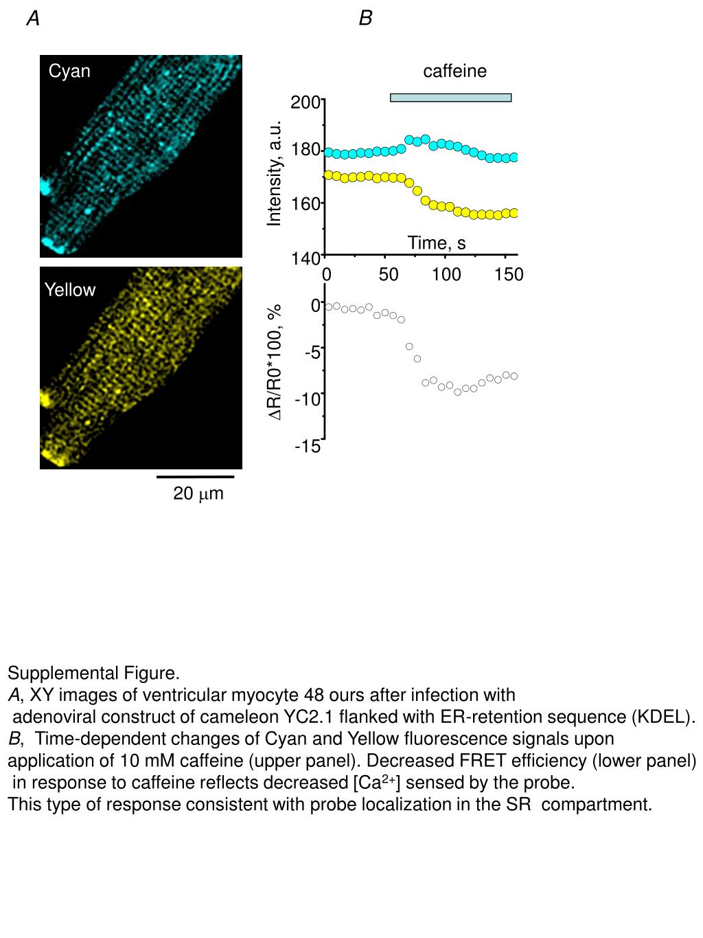
This study investigates the calcium dynamics in ventricular myocytes post-infection with an adenoviral construct of the calcium-sensitive probe Cameleon YC2.1. Fluorescent imaging reveals time-dependent changes in cyan and yellow fluorescence signals after administering 10 mM caffeine. Observations show a decreased FRET efficiency, indicating reduced intracellular calcium levels detected by the probe. These findings suggest the probe's localization within the sarcoplasmic reticulum (SR) and provide insights into cellular calcium signaling mechanisms in response to caffeine applications.
Télécharger la présentation 
Politique de téléchargement : le contenu du site vous est fourni TEL QUEL pour votre information et votre usage personnel ; il ne peut être vendu, concédé sous licence ni partagé sur d’autres sites sans l’accord de son auteur.Le contenu vous est fourni TEL QUEL, pour votre information et votre usage personnel uniquement. Téléchargez la présentation en cliquant sur ce lien. Lors du téléchargement, si vous ne parvenez pas à télécharger une présentation, l’éditeur a peut-être supprimé le fichier de son serveur.Pendant le téléchargement, si vous ne parvenez pas à obtenir une présentation, le fichier a peut-être été supprimé par l’éditeur.
F I N