Download
1 / 62
620 likes | 797 Vues
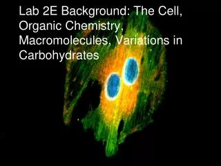
Lab 2E Background: The Cell, Organic Chemistry, Macromolecules, Variations in Carbohydrates. Cell Structures. What general features can be identified in this “typical” generalized cell?. A Tour Inside an Animal Cell. Cells. Structural unit of all living things

E N D
Lab 2E Background: The Cell, Organic Chemistry, Macromolecules, Variations in Carbohydrates
What general features can be identified in this “typical” generalized cell?
Cells • Structural unit of all living things • 50 – 100 trillion cells in human body • 200 different cell types that vary in size, shape, function • A cells SHAPE reflects its function • Plant Cells differ from animal cells by having: • Cell wall, chloroplasts, central vacuole, and tonoplast
Erythrocytes Fibroblasts Diversity of Cell Structure and Function Epithelial cells (a) Cells that connect body parts, form linings, or transport gases Skeletal muscle cell Nerve cell Smooth muscle cells (b) Cells that move organs and body parts (e) Cell that gathers information and controls body functions Fat cell Macrophage Sperm (c) Cell that stores nutrients (d) Cell that fights disease (f) Cell of reproduction
Nucleus • Porous phospholipid membrane • Inner membrane lined with intermediate filaments (nuclear lamina) that maintains shape • ER often is an extension of the nuclear membrane • Contain DNA of eukaryotic cells – “brain” of cell A single strand of DNA can be 3 meters long. How does all that DNA fit?
Condensation of Eukaryotic Chromosomes Nucleosome = DNA coils around histone proteins Chromatin = supercoiled nucleosomes Looped Domains = supercoiled chromatin Chromosome = supercoiled looped domains
Ribosomes • Assembles amino acids into polypeptide chain, which eventually folds into functional protein • Made of rRNA and protein • 2 subunits: large and small
Nucleolus • Located inside nucleus • makes ribosomal subunits by combining rRNA and proteins imported from cytoplasm • subunits leave nuclear pore and assembles into a ribosome in the cytoplasm
What is the endomembrane system? • System of membrane-bound organelles in cells that work cooperatively together to create secretory proteins, membrane-bound proteins, or plasma membrane proteins • Nucleus • ER • Golgi • Transport Vesicles • Lysosomes • Peroxisomes • Vacuoles • Plasma Membrane
Rough Endoplasmic Reticulum RER w/ bound ribosomes Space w/in ER = cisternae space Fcn: to fold and modify secretory proteins (glycoproteins) within cisternae space • attaches carbohydrates called oligosaccharides to growing and folding polypeptide chain • - vesicles bud off from RER and delivers glycoprotein to Golgi
Golgi Apparatus Adds “ID” tags (like phosphate groups) and uses these to “sort” proteins into different vesicles Dispatches vesicles w/glyco-proteins for shipping (trans side) Accepts vesicles from RER (cis side) Adds and removes monomers of sugar (small subunits) from glycoproteins
Rough ER Cisterna Proteins in cisterna Phagosome Membrane Vesicle Protein Synthesis and Export of Proteins Lysosomes containing acid hydrolase enzymes Vesicle incorporated into plasma membrane Pathway 3 Coatomer coat Golgi apparatus Pathway 2 Secretory vesicles Pathway 1 Plasma membrane Proteins Secretion by exocytosis Extracellular fluid
Stores water, organic compounds, ions, waste Supplemental role in endo and exocytosis as a “vesicle” Vacuoles
Lysosomes • Membrane-bound sac of digestive enzymes • Acidic env’t maintained by pumping H+ions from cytoplasm • Digests food, worn out cell parts, programmed cell death (webbing b/t fingers, tadpole tails)
Peroxisome • Breaks down toxic substances in liver • Breaks down fatty acids into carbohydrates for use in CR • In breakdown process, oxygen and hydrogen combine to create H2O2 • Peroxide = metabolic waste
Smooth ER • ER w/o ribosomes • Makes lipids, oils, steroids • Helps break down CHO • Detoxifies drugs by adding –OH groups water soluble toxins flushed from body
Mitochondria Cellular Respiration site requires oxygen (O2) to make ATP from glucose (C6H12O6) ATP is the energy form used for cellular work CO2 and H2O is produced as waste and bi-product of cellular respiration Mitochondria( The powerhouse of the cell!) Oxygen is delivered to our mitochondria from the air and carbon dioxide is released back as waste. Which system is responsible for this function?
Cytoskeleton Network of fibers in the cytoplasm that a) maintains cell shape/mechanical support b) anchors and/or moves organelles c) helps w/ cell motility 3 components 1) microtubules 2) microfilaments 3) intermediate filaments
Microtubules Structure: Hollow tube made up of α and β tubulin polypeptide 25 nm diameter Compression Resistent supports cell shape Forms spindle fibers for separation of chromosomes, makes up centrioles, and cilia/flagella
9 sets of 3 arrangement (ring formation) Ex. Centrioles, spindle fibers, basal body of cilia and flagella 9 + 2 arrangement (9 doublets surrounding a pair in the center) Ex. Cilia and Flagella Microtubule
Transmembrane/Integral protein Glycoproteins Glycolipid Phospholipids Cholesterol Peripheral/Surface Proteins Plasma Membrane Glycolipid and glycoproteins = Glycocalyx A cell boundary that selectively controls the movement of substances into and out of the cell = Selective Permeability Made up of a “mosaic” or collection
Why do cells need to increase permeability rate of the plasma membrane? Outside of cells bathed in interstitial fluid • Nutrient rich “soup” • Amino acids, sugars, fats, vitamins, hormones, proteins, salt, waste, neurotransmitters • Cells need to absorb what they need from this fluid AND remove waste in an efficient manner as needed One method: microvilli
Microvilli Fingerlike-extensions of plasma membrane Supported by actin (microfilament) core Microvilli and the glycocalyx help cells “stick” together (imagine your fingers interlocked and covered in sugar) Increases cell’s surface area relative to its volume, to increase absorptive and expelling properties
Cell Functions Molecular Transport • Two main categories of molecule transport exist in cells: active transport and passive transport • oxygen, ethanol, and carbon dioxide, can easily cross the membrane via passive transport, in the form of simple diffusion through a concentration gradient. • To transport macromolecules (proteins, lipids carbohydrates, nucleic acids), cells rely on active transport.
Cellular Respiration energy • When organisms release the _________ stored in carbohydrates and other food molecules Glucose Fructose Monosaccharides Sucrose: A disaccharide
Aerobic Respiration • Occurs when Oxygen is present • Many ATP Molecules made
Anaerobic Respiration • Occurs when no Oxygen is present • Very little ATP made • Two basic types: • Fermentation: Alcohol and CO2 produced • Lactic Acid
Reproduction • Mitosis • mostly used by somatic cells (cells of the body). • resulting daughter cell is an identical clone of the original cell • Meiosis • a form of sexual reproduction and only occurs in gametes (reproductive cells). • The resulting daughter cells have different genetic information than their parent cells.
Phases of Mitosis • Interphase: • Chromosomes are not clearly discerned in the nucleus, although a dark spot called the nucleolus may be visible. The cell may contain a pair of centrioles (or microtubule organizing centers in plants) both of which are organizational sites for microtubules.
2. Prophase: • Chromatin in the nucleus begins to condense and becomes visible in the light microscope as chromosomes. The nucleolus disappears. Centrioles begin moving to opposite ends of the cell and fibers extend from the centromeres. Some fibers cross the cell to form the mitotic spindle.
3. Prometaphase: • The nuclear membrane dissolves, marking the beginning of prometaphase. Proteins attach to the centromeres creating the kinetochores. Microtubules attach at the kinetochores and the chromosomes begin moving.
4. Metaphase: • Spindle fibers align the chromosomes along the middle of the cell nucleus. This line is referred to as the metaphase plate. This organization helps to ensure that in the next phase, when the chromosomes are separated, each new nucleus will receive one copy of each chromosome.
5. Anaphase: • The paired chromosomes separate at the kinetochores and move to opposite sides of the cell. Motion results from a combination of kinetochore movement along the spindle microtubules and through the physical interaction of polar microtubules.
6. Telophase: • Chromatids arrive at opposite poles of cell, and new membranes form around the daughter nuclei. The chromosomes disperse and are no longer visible under the light microscope. The spindle fibers disperse, and cytokinesis or the partitioning of the cell may also begin during this stage.
7. Cytokinesis • In animal cells, cytokinesis results when a fiber ring composed of a protein called actin around the center of the cell contracts pinching the cell into two daughter cells, each with one nucleus. In plant cells, the rigid wall requires that a cell plate be synthesized between the two daughter cells.
Race to the Board – Cell Drawing • Class divided into two teams to draw a cell, with all of its organelles. All organelles covered in lecture must be represented in the illustration. • No use of notes allowed. Must be done from memory. • All organelles must be accurately drawn, labeled with correct spelling. • Proper scientific illustration protocol must be followed.
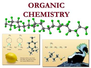
The Basics • CARBON (C): Always exists as having four bonds. An example of this is methane (see below). Notice that carbon has four bonds. • HYDROGEN (H): Always exists as having one bond. An example of this is water (see below). Notice that each hydrogen has one bond. • OXYGEN (O): Always exists as having two bonds and two lone pairs of electrons. An example of this can also be seen in water below. Notice that oxygen has two bonds and two lone pairs of electrons.
How To Calculate Formal Charge • Need periodic table • Notice that in the periodic table that all of the group numbers are listed across the top of the page. It is only the group numbers with the letters "a" next to them that we are concerned about.
Step One:Look up nitrogen (N) in the periodic table. Notice that it is in group 5(ignore the letter a). • Step Two: Count the number of covalent bonds (the lines) that are attached to Nitrogen. Notice that there are 4 lines attached to the nitrogen (N). • Step Three:Count the number of elctrons that are part of a lone pair (a lone pair looks like two dots). Notice that there are 0 electrons that are part of a lone pair on nitrogen. • Step Five:Now apply the formula which is • Group # - # of Covalent Bonds - # of lone electrons = Formal Charge • 5 - 4 -0= 1 • This means that the formal charge is 1 which means that nitrogen has a +1 charge.
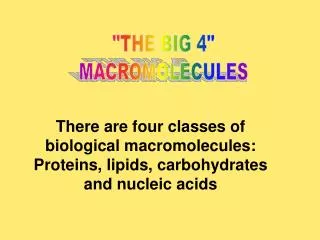
Proteins • made up from 20 different L-a-amino acids • A protein is a polypeptide, a linear polymer of many amino acids, linked by peptide bonds. • DNA codes for the amino acids that make up proteins through three letter strings of nucleotides called codons. • about 100,000 different proteins expressed in eukariotic systems • there are much fewer different structural motifs and folds, partly as a consequence of evolved pathways and mechansims
(Water is the byproduct of peptide bonds between amino acids)
Protein Functions Antibodies - are specialized proteins involved in defending the body from antigens (foreign invaders). One way antibodies destroy antigens is by immobilizing them so that they can be destroyed by white blood cells. Contractile Proteins - are responsible for movement. Examples include actin and myosin. These proteins are involved in muscle contraction and movement.
Enzymes - are proteins that facilitate biochemical reactions. They are often referred to as catalysts because they speed up chemical reactions. Examples include the enzymes lactase and pepsin. Lactase breaks down the sugar lactose found in milk. Pepsin is a digestive enzyme that works in the stomach to break down proteins in food. Hormonal Proteins- are messenger proteins which help to coordinate certain bodily activities. Examples include insulin, oxytocin, and somatotropin. Insulin regulates glucose metabolism by controlling the blood-sugar concentration. Oxytocin stimulates contractions in females during childbirth. Somatotropin is a growth hormone that stimulates protein production in muscle cells.
Structural Proteins- are fibrous and stringy and provide support. Examples include keratin, collagen, and elastin. Keratins strengthen protective coverings such as hair, quills, feathers, horns, and beaks. Collagens and elastin provide support for connective tissues such as tendons and ligaments.Storage Proteins- store amino acids. Examples include ovalbumin and casein. Ovalbumin is found in egg whites and casein is a milk-based protein.Transport Proteins- are carrier proteins which move molecules from one place to another around the body. Examples include hemoglobin and cytochromes. Hemoglobin transports oxygen through the blood. Cytochromes operate in the electron transport chain as electron carrier proteins.
Carbohydrates • Saccharides • Carbohydrates are used to store energy, though they serve other important functions as well. • The simplest carbohydrates are called monosaccharides, which has the basic structure (C·H2O)n, where n is three or greater. • Monosaccharides link together to form oligosaccharides and polysaccharides. Two monosaccharides link together to form a disaccharide.