
Data Collection Markers in Lumbar Spinal Segment: Defining Regions for Research
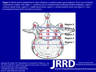
This illustration outlines the established marker points for data collection within sample lumbar spinal segments. Each row of markers represents distinct anatomical regions: Region 1 pertains to the posterior part of the vertebral body bordering the endplates, Region 2 is the anterior spinal canal, Region 3 denotes the middle spinal canal, Region 4 covers the posterior spinal canal, and Region 5 includes the area around facet joints and the spinous process. This methodology enhances the determination of the axial center of rotation in lumbar and thoracic spine segments.

Data Collection Markers in Lumbar Spinal Segment: Defining Regions for Research
E N D
Presentation Transcript
Figure 2. Marker points established for data collection in sample lumbar spinal segment. Each row of markers is defined by a region, with region 1 = posterior part of vertebral body bordering vertebral endplates, region 2 =anterior spinal canal, region 3 = middle spinal canal, region 4 = posteriorspinal canal, and region 5 = area encompassing facet joints and spinous process. Samagh SP, Rosen CD, Otarodifard K, Kornswiet M, Palmer G , Lee TQ. New method for determining apparent axial center of rotation of lumbar and thoracic spine segments.J Rehabil Res Dev. 2011;48(5):587-96.DOI:10.1682/JRRD.2010.09.0168