Download
1 / 11

E N D
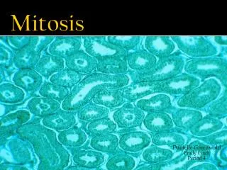
Prophase Chromatin in the nucleus begins to condense and becomes visible in the light microscope as chromosomes. The nucleolus disappears. Centrioles begin moving to opposite ends of the cell and fibers extend from the centromeres. Some fibers cross the cell to form the mitotic spindle. Prometaphase The nuclear membrane dissolves, marking the beginning of prometaphase. Proteins attach to the centromeres creating the kinetochores. Microtubules attach at the kinetochores and the chromosomes begin moving.
Metaphase Spindle fibers align the chromosomes along the middle of the cell nucleus. This line is referred to as the metaphase plate. This organization helps to ensure that in the next phase, when the chromosomes are separated, each new nucleus will receive one copy of each chromosome. Anaphase The paired chromosomes separate at the kinetochores and move to opposite sides of the cell. Motion results from a combination of kinetochore movement along the spindle microtubules and through the physical interaction of polar microtubules.
Telophase Chromatids arrive at opposite poles of cell, and new membranes form around the daughter nuclei. The chromosomes disperse and are no longer visible under the light microscope. The spindle fibers disperse, and cytokinesis or the partitioning of the cell may also begin during this stage. Cytokinesis In animal cells, cytokinesis results when a fiber ring composed of a protein called actin around the center of the cell contracts pinching the cell into two daughter cells, each with one nucleus. In plant cells, the rigid wall requires that a cell plate be synthesized between the two daughter cells.
Plant life cycles have two sequential phases that are termed alternation of generations : • The sporophyte phase is "diploid", and is that part of the life cycle in which meiosis occurs. However, many plant species are thought to arise by polyploidy, and the use of "diploid" in the last sentence was meant to indicate that the greater number of chromosome sets occur in this phase. • The gametophyte phase is "haploid", and is the part of the life cycle in which gametes are produced (by mitosis of haploid cells). In flowering plants (angiosperms) the multicelled visible plant (leaf, stem, etc.) is sporophyte, while pollen and ovaries contain the male and female gametophytes, respectively.
In preparation to switch from a mitotic tomeiotic sequence of events timing adjustmentsare made Mitosis Meiosis
Activities of meiosis that differ from mitosis • Pairing of homologous chromosomes • Crossing over between homologues • Reduction of chromosome number • Slow pace of meiotic prophase • Requirement of two cell divisionsinstead of one to complete the process • Lack of an S-period between the twodivisions