Download
1 / 85
850 likes | 1.07k Vues
Injuries to the Thigh, Leg, and Knee…. We will go over anatomy that covers bones, ligaments, tendons, muscles, nerves, and blood vessels of the region We will discuss the kinesiology of movements created by the muscles though the major joints

E N D
We will go over anatomy that covers bones, ligaments, tendons, muscles, nerves, and blood vessels of the region • We will discuss the kinesiology of movements created by the muscles though the major joints • The chapter continues with a description of soft-tissue injuries to the thigh that can become debilitating if not cared for properly • Including, contusions, strains, and various joint-related injuries
This chapter will cover issues such as osteochondritisdissecans, inflamed bursae, and patellar dislocation, along with injuries caused by chronic exercise • The chapter will also describe the four major ligaments of the knee and injuries to the knee joint that can be injured during sports participation • The chapter concludes with a discussion of prophylactic and functional knee bracing
Anatomy Review… • The lower extremity is an area where many athletes experience some type of injury during their sports career • The bones of the extremity include the femur, tibia, fibula, patella, and those of the foot (Gray, 1974) • http://www.youtube.com/watch?v=qoiaUV7fGEI
Anatomy Review… • The femur or thigh bone is the longest, strongest, and heaviest bone in the body • It has a rounded, ball-like head that attaches to the hip bone wit the help of ligaments • The head of the femur is attached to the shaft of the femur by a region known as the neck • The femur becomes flatter and wider as it proceeds toward the knee, where it articulates with the tibia
Anatomy Review… • The thigh has a great deal of blood and nerve tissue, both anteriorly and posteriorly • The anterior portion contains the long saphenous vein and several branches of the femoral nerve • The posterior section of the thigh are the deep femoral artery and the major nerve to the leg, the sciatic nerve • Most are quite well protected by the musculature of the thigh
Anatomy Review… • The muscles of the thigh can be broken down into three basic regions • First, the anterior muscles of the thigh, commonly called the quadriceps have two functions • The vastuslateralis, vastusintermedius, vastusmedialis and rectus femoris work together to extend the leg at the knee joint • Three of these muscles (VMO, intermedius, and lateralis) attach on the femur and run down the thigh to the quadriceps tendon
Anatomy Review… • The rectus femoris attaches on the hip bone at the anterior inferior iliac spine and runs down the leg to the quadriceps tendon • The other muscle in the anterior portion of the thigh is the satorius • It also attaches on the hip bone and runs somewhat diagonally down the thigh to the anterior medial portion of the tibialcondyle • This muscle is responsible for flexing, abduction, and lateral rotating the thigh at the hip
Anatomy Review… • The main muscles of the medial aspect of the thigh include the adductor longus, adductor brevis, adductor magnus and the gracilis • These muscles attach on the pelvis and run to the femur • The main function of these muscles is to adduct the hip with flexion of the thigh
Anatomy Review… • The third group of muscles in the thigh are in the posterior aspect of the thigh and are commonly known as the hamstrings • These include the semitendinosus, semimembranosus, and biceps femoris • All these muscles attach on the pelvis and run down the leg to the tibia • The main function of this group of muscles is to flex the leg at the knee
Anatomy Review… • http://www.youtube.com/watch?v=VdXAOWmbRuw
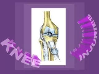
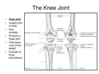
Anatomy Review… • The knee is a very complex joint • It can be damaged through any number of accidents occurring during sports participation • The femur and the tibia articulate with each other to form the tibiofemoral joint • The patella and the femur articulate with each other to form the patellofemoral joint
Anatomy Review… • The patella is a sesamoid bone, which means that it is totally enclosed within a tendon • In this case, the quadriceps tendon • The patella does not articulate with the tibia • Many ligaments support the knee joint
Anatomy Review… • There are 4 ligaments that serve as primary stabilizers of this joint: • Tibial or medial collateral ligament (MCL) • The fibular or lateral collateral ligament (LCL) • The anterior cruciate ligament (ACL) • Posterior cruciate ligament (PCL)
Anatomy Review… • The tibial (medial) collateral ligament extends from the medial epicondyle of the femur down to the medial condyle of the tibia • The tibular (lateral) collateral ligament begins at the lateral epicondyle of the femur and extends to the head of the fibula
Anatomy Review… • The fibular collateral ligament is the stronger of the two • Both ligaments help limit motion and/or disruption of the knee joint when movement at the joint is in a side-to-side direction • Valgus (knock-knees) • Varus (bow legs)
Anatomy Review… • The cruciate ligaments, unlike the collateral ligaments, are situated on the inside of the joint • The ACL attaches on the anterior portion of the intercondylar area of the tibia and runs superiorly and posteriorly to the internal aspect of the lateral femoral head
Anatomy Review… • The PCL attaches on the posterior aspect of the intercondylar area of the tibia and runs superiorly and anteriorly, passing the ACL on the medial side and attaching to the internal aspect of the medial femoral condyle • The function of these two ligaments is primarily to reduce or prevent anterior and posterior displacement of the femur or the tibia
Anatomy Review… • Two semicircular fibrocartilaginous disks, commonly called cartilage and more scientifically termed the menisci, are located within the space between the tibia and the femur • The menisci assist with the protection and nourishment of the knee joint, aid in the distribution of weight and stress applied to the joint surfaces, and help with the biomechanics of the joint
Anatomy Review… • There are two the medial and lateral menisci • Tendons of the muscles mentioned earlier in the description of the thigh run across the knee • Between the tendons and bone are several bursae, which reduce the friction of muscle tendons rubbing over a prominent area of bone, thereby adding some padding for the exposed bony areas of the knee
Common Sports Injuries • Injuries can occur in any sport • This area can sustain injuries that are a result of overuse, trauma caused by an opponent, or trauma produced by the power and explosive movements required in some sports • Because the knee is part of a complex mechanical system that includes the foot, ankle, lower leg, hip, and pelvis, there are times when another part of this system causes problem that can eventually be exhibited in the knee
Skeletal Injuries…Femoral Fractures • The femur is the longest bone ins the body and is therefore subject to being fractured • However, this requires a great deal of force and is not common occurrence in sports • If a fracture does occur to the shaft of the femur as a result of sports participation, the injury is quite obvious • The athlete should not attempt to walk on a femoral fracture
Skeletal Injuries…Femoral Fractures • The athlete must be immediately transported to the nearest medical facility with the leg splinted and without bearing any weight on the affected limb • A femoral fracture requires urgent medical attention because the initial trauma can lead to multiple problems • Including a lack of circulation, nervous innervations, or shock and other medical issues
Skeletal Injuries…Femoral Fractures • The neck of the femur can also be fractured • This occurs more often in sports than does a fracture of the shaft • Older children and teenagers are at greater risk for this injury because the fracture can potentially occur at the site of a growth plate • Among younger athletes these fractures can be the result of direct trauma or overuse
Skeletal Injuries…Femoral Fractures • If direct trauma is the cause, the athlete typically had a foot planted and then got hit in the hip or upper thigh with a great deal of force • This injury needs to be evaluated ASAP by a physician • Once complication of a fracture in the neck of the femur is avascular necrosis (tissue death) of the femoral head • Caused by a blood supply to the bony portion of the femoral head
Skeletal Injuries…Femoral Fractures • S&S • Pain at the site of injury • Difficulty ambulating on the affected leg • Swelling and/or deformity may occur • Athlete may report a traumatic event as the cause • The athlete may report having heard or felt a severe pop or snap at the time of injury • TX: • Be prepared to treat the athlete for shock if necessary • Splint the injured leg, preferably with a traction splint • Apply sterile dressings to any related open wounds • Monitor vital signs and circulation to the lower leg • Arrange for transport to the nearest medical facility
Skeletal Injuries…Patella Fracture • Other skeletal problems include a fracture of the patella and dislocation of the knee or tibiofemoral joint • Although the patella can be fractured, this is not a common occurrence in sports • A patellar fracture is caused by violent trauma, and the athlete is incapacitated for a short period of time • There is a great deal of pain associated with this injury
Skeletal Injuries…dislocation of tibiofemoral joint • A dislocation can sometimes compromise the blood flow to the lower leg • If there is a dislocation of the tibiofemoral joint, this is outwardly apparent, and the athlete will experience marked pain • Must be splinted, and the athlete must be referred to the nearest medical facility without delay • http://www.youtube.com/watch?v=-kRMSYelGTU
Soft-tissue Injuries to the thigh • Most of the soft-tissue injuries to the thigh are either the result of contact with an opponent or explosive movement by the athlete causing a self-inflicted muscle strain • Many sports, such as football and hockey use some type of protective padding to prevent contact • However, complete prevention is not always possible, and injuries do occur
MyositisOssificans… • When an athlete receives a blow to the quadriceps muscle group, there is a contusion to the musculature from some other violent force (internally or externally), bleeding and damage often occur within the muscle fibers • Depending on the force of impact and the muscles involved, the contusion may be of varying degrees of severity • In any case, the athlete must be counseled about the care of this injury and the long term complications of improper care of a muscular contusion
MyositisOssificans… • The initial muscular contusion causes bleeding • If not cared for properly, or if further damage occurs, there is an increase in the amount of blood lost in the same area • Over a long period of time, continued bleeding and insult to the area can result in calcification within the muscle, abnormal bone growth, and further dissability (Larson et al., 2002)
MyositisOssificans… S&S: TX: • The athlete will report a forceful impact to the area • Muscular tightness and swelling may be present • Athlete has decreased ability to forcefully contract the muscle • Athlete has difficulties in ambulating with the affected leg • Apply ice and compression immediately • If the injury is severe, place the athlete on crutches • Have the athlete rest and avoid any contact with the area • The athlete must be allowed plenty of rest and time • Early controlled movement of the controlled contused muscle assist in regenerating the muscle
MyositisOssificans… • The early mobilization in this case must be well controlled • The athlete should not be allowed to participate in full contact practice or competition until complete healing has occurred • The area should be padded if the athlete continues to participate • Moreover, the athlete should be well aware of the long-term consequences of continued trauma to the area
Muscular Strains to the Thigh… • Most of the strains to athletes, however, are to the hamstrings and adductor muscles • Strains to the adductor muscles are commonly known as groin pulls • Most strains occur to the muscle itself and not the tendon • Such strains are usually the result of muscles being stretchered too far, which is the case with the adductor muscles
Muscular Strains to the Thigh… • However, strains can be the result of miscommunication between agonistic muscles and antagonistic muscles • Agonistic muscle, muscles in a state of contraction as related to opposing muscles • Antagonistic muscle, muscles that counteract the action of agonistic muscles • http://www.youtube.com/watch?v=i2VG3HGBrBw
Muscular Strains to the Thigh… • If the muscle is stretched too far, the fibers of the muscle are damaged and bleeding occurs • Which leads to loss of contractibility, stiffness, and impaired movement • In conjunction with agonistic and antagonistic muscles, the quadriceps musculature is contracting while the hamstrings are also contracting, causing the weaker muscle to be torn and damaged • Typically the hamstrings are the weaker of the two groups • Therefore, this is the musculature that is usually strained
Muscular Strains to the Thigh… • Many athletes experience chronic tightness and repetitive strains to the muscles of the thigh adductor (groin) region • Specifically the adductor brevis, longus, and magnus muscles can exhibit problems • Especially in athletes participating in activities requiring multiple changes in speed and/or direction • Is it not uncommon for a track, soccer, football, or volleyball athlete to c/o tight, sore, or strained muscles
Muscular Strains to the Thigh… • The groin muscles are critical movers in speed and change of direction movements and are not easy to warm up and stretch • Special attention must be given
Muscular Strains to the Thigh… • These groin muscles can be debilitating if not cared for properly and quickly • Typically, when a strain to one or more of the groin muscles occurs, the athlete feels a sharp pain in the medial side of the thigh, possibility associated with a “tearing” feeling • Not long after the incident the athlete will c/o soreness, stiffness, and a lack of movement in the area
Muscular Strains to the Thigh… • During the recovery, athletes need to implement a stretching program that specifically targets the adductor muscles • Stretching must be an integral part of the recovery from this and any other muscle strain injury because of the need to reduce scarring of the affected muscles • http://www.youtube.com/watch?v=ZY9LWbqEz5Q
Muscular Strains to the Thigh… S&S TX: • A sharp pain in the affected muscle • Swelling and inflammation in the immediate area • Weakness and inability of the muscle to contract • After a few days there may be discoloration of the area • In severe cases, a visible defect is noted in the muscle • Apply ice and compression immediately • Have the athlete rest and use crutches if necessary • Have the athlete evaluated by a member of the medical team
Patellofemoral Joint Injuries… • Several injuries to the patellofemoral joint, both chronic and acute, can become debilitating • Intervention is required if the athlete is to return to participation at peak level • Some of the problems causing injury are the result of faulty mechanics or growth in adolescents and are not caused by anything that could be prevented initially
OsteochondritisDissecans • Also called “joint mice” because small pieces of bone that have been dislodged or chipped from the joint are floating within the joint capsule • In adolescents, OCD is the most common cause of a loose body in the joint space (Hixon & Gibbs, 2000) • This can lead to serious problems • When the joint surfaces are damaged and no longer make smooth contact with each other, further pain and joint damage are almost always inevitable
OsteochondritisDissecans • The piece of bone does not always have to be freely floating within the joint space • It may be dislodged yet still attached to the original bone and causing painful movement • If in fact the piece of bone is freely floating within the joint space, it can cause a blocking or locking action that limits the movement at the knee joint • May juvenile athletes respond to conservative treatment, whereas others may require surgical intervention • http://www.youtube.com/watch?v=of0gg-zXERA
OsteochondritisDissecans S&S TX: • Chronic knee pain with exertion that is generalized, not specific • There may be chronic swelling present • The knee may lock if there is a loose body within the joint • The athlete may be unable to fully extend the extremity • The quadriceps group may atrophy (lose muscle tone) • One or both femoral condyles may be tender to palpation when the knee is flexed • Apply ice and compression • If the athlete has difficulty walking or the knee is locking, have the player use crutches • Have the athlete use a physician for proper treatment
Inflamed Bursae • A bursa is a small fluid-filled sac located at a strategic point in the body that assist in the prevention of friction between bony surfaces, tendons, muscles, or skin • There are numerous bursae in the knee joint • However, only a few are commonly irritated • A bursa can become inflamed as a result of trauma or infection • Can also be due to chronic overuse and irritation of the bursa
Inflamed Bursae • The prepatellar bursa is located just under the skin and above the patella and can be susceptible to direct trauma • The constant use of the legs and knees in some exercises creates too much friction in the area, and the bursae respond by becoming inflamed from direct trauma
Inflamed Bursae S&S: TX: • Swelling and tenderness at the site • Increased pressure externally typically causes pain • The athlete may report direct trauma or a chronic buildup of swelling • Apply ice and compression • Reduce activity for a short period of time • In chronic cases, anti-inflammatory agents may be helpful
Patellar Dislocation/Subluxation • When an athlete makes a quick, cutting motion to one side or another, a great deal of abnormal force is generated within the knee • As a result of this sudden abnormal force, the patella can move laterally instead of superiorly and inferiorly as it normally does • If the patella moves too far laterally, it can become dislocated (subluxation) • http://www.youtube.com/watch?v=qbFgl5iL_zw