Download
1 / 28
310 likes | 919 Vues
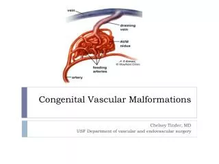
Department of Neurological Surgery. Cavernous Malformations, Venous Malformations, and Capillary Telangiectasias. Richard E. Clatterbuck, M.D., Ph.D. The Johns Hopkins University School of Medicine. Cavernous Malformations.

E N D
Department of Neurological Surgery Cavernous Malformations, Venous Malformations, and Capillary Telangiectasias Richard E. Clatterbuck, M.D., Ph.D. The Johns Hopkins University School of Medicine
Cavernous Malformations • Abnormal vascular channels lined by endothelial cells (cells that line all blood vessels) but lacking other components of typical blood vessel walls • Have been described as having an appearance similar to mulberries
AKA • Cavernous angioma • Cavernous hemangioma • Cerebral angioma • Cavernoma • Cavmals • CCMs (cerebral cavernous malformations)
Radiographic Appearance • Classically described as a “popcorn” lesion with a reticulated appearance on MRI scans
Epidemiology • Occur in 0.5% of the population (1 in 200 people) and may represent 10% of vascular malformations • Perhaps slightly more prevalent in females, 1.8:1 in our series
Presentation • Mean age 34.6 years (7.8-78.5) • Average of 3.4 lesions per patient, solitary in 63% and multiple in 25% • 19% had venous malformations • 4% had capillary telangiectasias • Headache (65%), seizures (49%), focal deficits (46%), hemorrhage (13%), asymptomatic (1.5%)
Hemorrhage rates • 3.1% per patient year hemorrhage rate • 0.9% in males • 4.2% in females • No difference in lesions in the cerebrum or brainstem
Seizure rates • 4.8% per patient year • 2.4% new onset seizure rate per patient year • 5.5% recurrent seizure rate per patient year
Venous malformation • Persistence of a developmentally expressed venous drainage pattern • Classically described as a caput medusae appearance
AKA • Developmental venous anomaly (DVA) • Venous anomaly • Venous angioma • Venous malformation
Presentation • Mean age 39.1 years (18.7-73) • 19% had another cerebrovascular malformations • Headache (50.8%), seizures (30.2%)
Hemorrhage rate • 0.15% per lesion year
Capillary Telangiectasia • Normal capillary structurally at the cellular level but abnormal in size, dramatically dilated
AKA • Capillary malformations • Captels
Presentation • Much rarer lesions with unclear although certainly low hemorrhage rate • Present throughout life but typically in 3rd or 4th decade • Symptoms include headache, numbness, dizziness, visual disturbance
Cerebral Vascular Malformations • Cavernous malformation • Venous malformation • Capillary telangiectasia