Download
1 / 42
440 likes | 1.08k Vues
Approach to the Mediastinum in Trauma:. Density vs. Width Tammy Washut MS4. Traumatic Injuries to Worry About. Mediastinal hematoma Aorta/great vessel injury Spinal hematoma. Rapid deceleration injury Blunt chest trauma- MVA, falls. The sudden stop causes the blood filled descending

E N D
Approach to the Mediastinum in Trauma: Density vs. Width Tammy Washut MS4
Traumatic Injuries to Worry About • Mediastinal hematoma • Aorta/great vessel injury • Spinal hematoma
Rapid deceleration injury Blunt chest trauma- MVA, falls The sudden stop causes the blood filled descending aorta to “snap”. The aortic arch is fixed in position by branches from the arch. Mechanism Sudden deceleration as sternum hits steering wheel. As the aortic tube “snaps”, the intima is torn just distal to left sub- clavian artery.
External signs • “Seat belt sign” • Chest ecchymosis • Sternal/Rib fractures
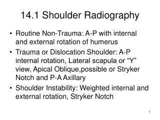
Chest X-ray • Classically taught to look for widened mediastinum • Wide mediastinum = 8 cm • What is the problem with this??? • Wide mediastinum has a broader differential than paratracheal density
Causes of Wide Mediastinum • Magnification • Rotation • Mediastinal hematoma • Spinal hematoma • Lymphadenopathy • Long intravascular volume • Obese patients
Magnification • Film placed directly behind the patient • Initially used to determine the 8 cm criteria for wide mediastinum
Magnification • Film placed under the backboard • 17% enlargement
Magnification • Film placed in trauma bed • 25% enlargement
Rotation 4 cm 7 cm Rotated Right Rotated Left
7 cm 4 cm Intravascular Volume Pre-Dialysis Post-Dialysis
Right Paratracheal Density • Composed of azygous vein and SVC • Density normally less than aortic arch • Increased = hematoma • Why? • Not affected by technical factors • Simple
Right Paratracheal Density Normal Increased Density
Mediastinal Hematoma • Other Signs: • PT stripe • Apical cap • Aortic Arch • NG deviation • Tracheal deviation
Mediastinal Hematoma • Other Signs: • PT stripe • Apical cap • Aortic Arch • NG deviation • Tracheal deviation
Mediastinal Hematoma • Other Signs: • PT stripe • Apical cap • Aortic Arch • NG deviation • Tracheal deviation
Mediastinal Hematoma • Other Signs: • PT stripe • Apical cap • Aortic Arch • NG deviation • Tracheal deviation
Mediastinal Hematoma • Other Signs: • PT stripe • Apical cap • Aortic Arch • NG deviation • Tracheal deviation
Hypothesis: OHSU DATA Right paratracheal density is discriminatory sign in trauma patients with widened mediastinum
Methods • 122 Trauma patients (2001-2003) • Screening Trauma chest radiograph • Mediastinal width > 8.0 cm • CT Chest w/contrast within 24 hours • Four readers of different levels of training • R paratracheal region evaluated
Methods • Patients categorized by ISS • AIS by body region • Chest: 1-6 • Low risk: 0-2 (80 patients) • High risk: >2 (42 patients)
Results • 19 mediastinal hematomas (15.6%) • 13 high-risk • 6 low-risk • 5 aortic injuries (4.1%) • 4 deceased (3.3%)
Results Sensitivity
Limitations • Single institution • Presented to readers in artificial setting • Relatively few hematomas • AIS/ISS scoring not useful as triage tool
Strengths • Trauma patients with widened mediastinum • Confirmed by CT w/in 24 hours • Blinded analysis • Clinical information available on all patients
Conclusions • Screening chest radiograph valuable in low/moderate risk trauma patients • Right paratracheal density valuable • Avoid CT in low-risk patients • 7.3% normal mediastinum • High risk patients should have CT
Recommendations • Low risk patients with or without a wide mediastinum but no paratracheal density do not need to have CT of the chest • High risk patients with mechanism of injury (i.e. seatbelt sign) should go to CT regardless • Paratracheal density, not width, should direct further management
Possible Hematoma? • Yes- aortic rupture • Paratracheal density on right and loss of aortic arch definition
Possible Hematoma? • Yes, but in this case it is lymphadenopathy in a high risk trauma patient • This patient should get a chest CT
Possible Hematoma? • No- mediastinum is wide, but no paratracheal density • Patient is rotated to right
References • Melton SM et al., J Trauma 2004; 56:243-250 • Mirvis SE et al., Radiology 1987; 13:487-493 • Baker SP et al., J Trauma 1974; 14:187-196 • Woodring JH et al., Radiology 1984; 151:15-21 • Parmley LF et al., Circulation 1953; 17:1086-1101 • Woodring JH et al., J Emerg Med 1990; 8:467-476 • Woodring D et al., Ann Thor Surg 1984; 37:171-178 • Blackmore CC et al., Emerg Radiology 2000; 7:142-148 • Patel NH et al., Radiology 1998; 209:335-348 • Milne EN et al., Radiology 1984; 153:25-31 • Demetriades D et al., Arch Surg 1998; 133:1084-1088 • O’Connor CE et al., Emerg Med J 2004; 21:414-419 • Special thanks to Dr. Marc Gosselin and Dr Peter Verhey for references, • images and study data and slides.