Download

1 / 37
450 likes | 1.38k Vues
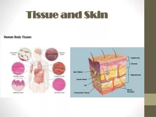
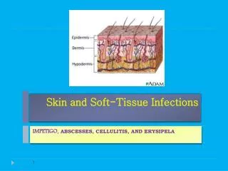
SKIN AND SUBCUTANEOUS TISSUE. I. Introduction A. Function 1. Protection 2. Thermoregulation 3. Sensory. B. Anatomy 1. Epidermis – most cellular layer a. keratinocytes – most numerous and forms a mechanical barrier b.Langerhan’s – immunologic function

E N D
I. Introduction A. Function 1. Protection 2. Thermoregulation 3. Sensory
B. Anatomy 1. Epidermis – most cellular layer a. keratinocytes – most numerous and forms a mechanical barrier b.Langerhan’s – immunologic function c. Melanocytes – pigment
2. Dermis – supporting layer, mostly fibroblast which produce collagen 3. Basement layer – dermal epidermal junction - first layer where blood vessel and lymphatics are present - if lesion has not crossed this layer, it is called an “in-situ” lesion
II. Pathology A. Trauma 1. Dirty and infected wounds – debridement and closed by secondary intention 2. Lacerations – closed primarily
B. Decubitus Ulcer or Pressure Ulcer - excessive, unrelieved pressure (60 cm Hg applied for 1 hour) - muscle more sensitive than skin to ischemia - Tx. – debridement and grafting
C. Keloid and Hypetrophic Scar - over abundance of deposition of collagen 1. Hypertrophic scar – nodularity remains within the incision - no treatment necessary 2. Keloid – nodularity goes beyond the incision - seen more in children and across sternum - treated with triamcinolone
D. Infections 1. Folliculitis – infected hair follicle - caused by Staph. sp. - leads to furuncle carbuncle - Tx. – incision and drainage and antibiotics 2. Hidradenitis suppuritiva - plugged apocrine gland in axilla and inguinal area - Tx. – warm compress, hygiene, discontinuation of deodorants, open drainage if recurrent
3. Pilonidal disease – infected pilosebaceous cysts in the saccrococygeal area, lined by granulation tissue - Tx. – drainage, currete
4. Staphyloccocal Scalded Skin Syndrome - erythema, bullae formation, loss of epidermis - caused by exotoxin from staphyloccocal infection - similar to partial thickness burn -cleavage is in the granular layer - Tx. – replace fluid, electrolytes, skin care, antibiotics
5. Toxic Epidermal Necrolysis - Immunologic reaction to certain drugs such as sulfonamides, phenytoin, barbituates, and tetracycline - Tx. – same as SSSS 6. Viral – verruca vulgaris, associated with pappiloma virus - associated with squamous cell ca - Tx. – chemical, electrocautery, surgery
E. Benign Tumors Cysts 1. epidermal – sebaceous cysts, most common 2. Trichilemmal – occurs more commonly in females 3. Dermoid – results from epithelium trapped during midline closure in fetal development - Tx. - excision
F. Nevi 1. Acquired a. Junctional – epidermis b. Compound – migrates partially down to the dermis c. Dermal – cells at dermal layer - involutes
2. Congenital – rare - large and may contain hair - occurs in bathing trunks distribution - Tx. - excision
G. Vascular 1. Hemangioma a. capillary (strawberry) - compressible, vascular lesion with sharp borders - located mostly in the face, scalp, and shoulder - observe, 90% involute
b. Cavernous - bright red or purple, with spongy consistency - Tx. – excision 2. Vascular malformation - enlarged vascular spaces lined with non proliferating endothelial cells a. portwine stain – capillary malformation - Tx. – embolization b. glomus tumor – painful blue –gray nodules - arises from the glomus body or Sucquet- Hoyer canal found in the dermis and contributes to thermal regulation - may lead to glomangiosarcoma - Tx. - excision
H. Soft Tissue Tumors ( achrocordons, lipomas, dermatofibromas) - Tx. – excision I. Neural - Neurofibromas (café-au-lait spots) - associated with von Reklinghausen’s disease
J. Malignant Tumors 1. Epidemiology a. malignant radiation b. chemicals c. viral d. chronic irritation e. immunosuppresion
2. Types a. basal cell carcinoma - most common - slow growing, rare metastases - excision with 2-4 mm margin
b. squamous cell carcinoma - metastasizes faster - Bowen’s disease – ca-in-situ - Erythroplasia of Queyrat – ca of the penis - lesion more than 1 cm has 50% chance of metastasis - Tx. – excision with 1 cm margin - Moh’s technique – serial excision to preserve skin
c. malignant melanoma - arises from dysplastic melanocytes i. superficial spreading - most common (70%) - flat with areas of regression
ii. nodular – 15-20% - dark, slightly raised - growth more vertical than radial iii. lentigo malignant 5-10% - best prognosis - occurs in areas of high solar degeneration
b. prognostication i. Clark ii. Breslow iii other factors - anatomic location – extremities better than trunk or face - ulceration
- inflammatory infitrates - sex - histologic type c. treatment - still primarily surgical i. in-situ - .5 to 1 cm margin ii. T1 (smaller than .76 mm) - 1-2 cm iii. thicker lesion – 3 cm margin - excision is up to the deep fascia - chemotherapy - palpable nodes are removed by regional dissection