Download
1 / 4
40 likes | 168 Vues
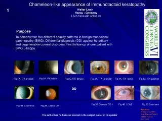
Chameleon-like appearance of immunotactoid keratopathy. 1. Walter Lisch Hanau ; Germany. Lisch.hanau@t-online.de. Purpose. To demonstrate five different opacity patterns in benign monoclonal gammopathy (BMG). Differential diagnosis (DD) against hereditary

E N D
Chameleon-like appearance of immunotactoid keratopathy 1 Walter Lisch Hanau ; Germany Lisch.hanau@t-online.de Purpose To demonstrate five different opacity patterns in benign monoclonal gammopathy (BMG). Differential diagnosis (DD) against hereditary and degenerative corneal disorders. First follow-up of one patient with BMG L-kappa. Fig.2A. ITK-lattice Fig.1A. ITK-crystals Fig.2C. ITK-diffuse Fig. 3A. ITK- granular Fig.4A. ITK- band Fig.5A. ITK-patches . DD Fig.3B.Granular CD 1 Fig.4B. LCAT Fig.5B.Salzmann Fig.1B. Cystinosis Fig.2B. Lattice CD Address : Walter Lisch MD Kurt-Blaum-Platz 8 63450 Hanau Germany The author has no financial interest in the subject matter of this poster
Chameleon-like appearance of immunotactoid keratopathy 2 Walter Lisch Hanau ; Germany Lisch.hanau@t-online.de Methods Colored slit-lamp photo-documentation of five BGM L-kappa patients with different types of immunotactoid keratopathy (ITK) : Pat. 1 : Classical crystalline-like ITK (Fig.1A); Pat. 2 : Central lattice-like ITK in 1992 (Fig. 2A ) and moderate diffuse-like ITK in 2009 (Fig.2C); Pat. 3 : Peripheral granular-like ITK (Fig 3A); Pat. 4 : Peripheral band-like ITK (Fig.4A); Pat. 5 : Peripheral patches-like ITK (Fig.5A). Systemic and serological examination of all five patients. Fig.2A. ITK-lattice Fig.1A. ITK-crystals Fig.2C. ITK-diffuse Fig. 3A. ITK- granular Fig.4A. ITK- band Fig.5A. ITK-patches . DD Fig.3B.Granular CD 1 Fig.4B. LCAT Fig.5B.Salzmann Fig.1B. Cystinosis Fig.2B. Lattice CD Address : Walter Lisch MD Kurt-Blaum-Platz 8 63450 Hanau Germany The author has no financial interest in the subject matter of this poster
Chameleon-like appearance of immunotactoid keratopathy 3 Walter Lisch Hanau ; Germany Lisch.hanau@t-online.de Results The systemic and serologic examination disclose in all five patients a BMG L-kappa.The 17-year follow-up of Pat.2 shows a change and reduction of the corneal opacities due to a strict lower protein nutrition (Fig.2A and 2C).Differential diagnosis : Pat.1 against cystinosis (Fig.1B) and Schnyder; Pat.2 against lattice CD (Fig.2B); Pat.3 against granular CD 1 (Fig.3B); Pat.4 against arcus lipoides and LCAT (Fig.4B); Pat.5 against gelatinous CD and Salzmann (Fig.5B). Fig.2A. ITK-lattice Fig.1A. ITK-crystals Fig.2C. ITK-diffuse Fig. 3A. ITK- granular Fig.4A. ITK- band Fig.5A. ITK-patches . DD Fig.3B.Granular CD 1 Fig.4B. LCAT Fig.5B.Salzmann Fig.1B. Cystinosis Fig.2B. Lattice CD Address : Walter Lisch MD Kurt-Blaum-Platz 8 63450 Hanau Germany The author has no financial interest in the subject matter of this poster
Chameleon-like appearance of immunotactoid keratopathy 4 Walter Lisch Hanau ; Germany Lisch.hanau@t-online.de Conclusions ITK can mimick some forms of hereditary and degenerative corneal disorders(1). ITK can be the first symptom of BGM. An annual internal check of BGM is to recommend because of the occurrence of malign MG in 20% of cases(2). References 1) Garibaldi DC et al. Surv Ophthalmol. 2005;50:61-80 2) Spiegel P et al. Cornea. 1990;9/1:81-85 Fig.2A. ITK-lattice Fig.1A. ITK-crystals Fig.2C. ITK-diffuse Fig. 3A. ITK- granular Fig.4A. ITK- band Fig.5A. ITK-patches . DD Fig.3B.Granular CD 1 Fig.4B. LCAT Fig.5B.Salzmann Fig.1B. Cystinosis Fig.2B. Lattice CD Address : Walter Lisch MD Kurt-Blaum-Platz 8 63450 Hanau Germany The author has no financial interest in the subject matter of this poster