Myelodysplastic Syndromes
750 likes | 1.7k Vues
Myelodysplastic Syndromes. Nicole N. Balmer M.D. June 3 rd , 2005. History of MDS. First described in 1938 - 100 patients with refractory anemia were described; Subsequently, the terms "preleukemic anemia“ and “preleukemia” were used.

Myelodysplastic Syndromes
E N D
Presentation Transcript
Myelodysplastic Syndromes Nicole N. Balmer M.D. June 3rd, 2005
History of MDS • First described in 1938 - 100 patients with refractory anemia were described; Subsequently, the terms "preleukemic anemia“ and “preleukemia” were used. • In 1963, a variant of acute leukemia was described, characterized by a prolonged and often benign clinical course, with a comparatively lower but variable percentage of bone marrow blasts; the authors termed this condition "smoldering acute leukemia"
History of MDS • In the 1970s, chronic myelomonocytic leukemia (CMML) was recognized as a unique preleukemic syndrome. • In 1976, the French-American-British (FAB) Cooperative Group initially defined refractory anemia with excess blasts (RAEB) and CMML as preleukemic states. Six years later, the FAB group added three more categories to this classification scheme and adopted the present term "myelodysplastic syndromes". • These disorders, and other members of the MDS "family“ were subsequently defined by the WHO.
The Myelodyplastic Syndromes • Six types of myelodysplastic syndromes according to WHO. • Refractory anemia • Refractory anemia with ringed sideroblasts • Refractory cytopenia with multilineage dysplasia • Refractory anemia with excess blasts • Myelodysplastic syndrome, unclassifiable • 5q- syndrome (myelodysplastic syndrome associated with isolated del (5q) chromosome abnormality • Related syndromes: • Myelodysplastic/Myeloproliferative diseases
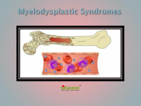
Myelodysplastic Syndromes • MDS Definition: • A group of disorders presenting with some evidence of bone marrow failure and dysplasia of one or more of the myeloid lineages, with <20% blasts in the blood or marrow. • Epidemiology: • Occur primarily in older patients (most common > 70 years).
MDS – Clinical Symptoms • Ecchymoses • Fatigue • Pallor • Ecchymoses/petechiae • Abnormal bleeding • Infection
MDS Etiology • Two etiologic categories of MDS: 1.) De Novo: Associated with: -benzene exposure (gasoline) -cigarettesmoking -viruses -Fanconi’s anemia 2.) Therapy related: Associated with: -alkylating agent chemotherapy -radiation
Prognostic Groups • Two groups based on survival and evolution to acute leukemia • 1.) “Good” group • Refractory anemia (RA) • Refractory anemia with ringed sideroblasts (RARS) • 5q - syndrome • 2.) “Bad” group • Refractory anemia with excess blasts (RAEB) • Refractory cytopenia with multilineage dysplasia (RCMD) • MDS unclassified can be either
Prognostic Scoring • The International Myelodysplastic Syndrome Working Group developed a scoring system based on 3 variables:
International Prognostic Scoring System Data (IPSS) • Overall median survival was 5.7, 3.5, 1.2, and 0.4 years for patients with IPSS scores of zero (low risk), 0.5 to 1.0 (intermediate-1 risk), 1.5 to 2.0 (intermediate-2 risk), and 2.5 to 3.5 (high risk), respectively. The time for 25 percent of the patients in each of the four risk groups to evolve into acute leukemia was 9.4, 3.3, 1.1, and 0.2 years, respectively.
IPSS • Other adverse prognostic factors which may improve the prognostic value of the IPSS include: • -CD34 positivity of bone marrow nucleated cells -Increased expression of the Wilms' tumor gene (WT1) -Increased serum beta-2 microglobulin concentration -Mutations of the FLT3 gene -Abnormal localization of immature precursors (ALIP).
Refractory Anemia • RA Definition: • Dyplasia of the erythroid series only. • Clinically, anemia is refractory to hematinic therapy • Myeloblasts < 1% blood and < 5% marrow • <15% ringed sideroblasts in marrow • No Auer rods • Other etiologies of erythroid abnormalities must be excluded. These include: • drug/toxin exposure -vitamin deficiency • viral infection -congenital disease
Refractory Anemia • Epidemiology: • 5-10% of MDS cases. • Older patients • Morphology: • Anisopoikilocytosis on peripheral smears • Dyserythropoiesis with nuclear abnormalities (megaloblastoid change) • < 15% ringed sideroblasts
Refractory Anemia • Genetics: • 25% may have genetic abnormalities • Prognosis: • Median survival is 66 months • 6% rate of progression to acute leukemia
Refractory Anemia with Ringed Sideroblasts • RARS definition: • Dyplasia of the erythroid series only. • Clinically, anemia is refractory to hematinic therapy • Myeloblasts < 5% in marrow, absent in blood • >15% ringed sideroblasts in marrow • No Auer rods • Other etiologies of ringed sideroblasts must be excluded. These include: • Anti- tuberculosis drugs • Alcoholism
Refractory Anemia with Ringed Sideroblasts • Epidemiology: • 10-12% of MDS cases. • Older patients • Males > females • Morphology: • Dimorphic pattern on peripheral smears • Majority RBC’s normochromic, 2nd population hypochromic • Dyserythropoiesis with nuclear abnormalities (megaloblastoid change)
Refractory Anemia with Ringed Sideroblasts • Morphology (con’t.) • < 15% ringed sideroblasts (RS) • RS = Erythroid precursor with ≥ 10 siderotic granules encircling 1/3 or more of the nucleus. • If excess blasts present, this dictates diagnosis, despite percentage of RS’s.
Refractory Anemia with Ringed Sideroblasts • Genetics: • Clonal chromosomal abnormalities in • <10%; in fact, development of such an abnormality should prompt reassessment of diagnosis. • Prognosis: • Median survival 6 years (72 months) • 1-2% rate of progression to acute leukemia
Refractory Cytopenia with Multilineage Dysplasia • RCMD definition: • Dyplasia in 10% or more of cells in 2 or more myeloid lines. • Myeloblasts < 1% blasts in the blood and < 5% in marrow. • No Auer rods • < 1 x 10⁹/L monocytes in blood
Refractory Cytopenia with Multilineage Dysplasia • Epidemiology: • 24% of MDS cases. • Older patients Morphology: • Neutrophil abnormalities may include: • Hypogranulation • Pseudo-Pelger-huet (hyposegmentation/barbells) • Megkaryocyte abnormalities may include • Hypolobation -Micromegakaryocytes
Refractory Cytopenia with Multilineage Dysplasia • Morphology (con’t.) • Erythroid abnormalities may include nuclear abnormalities such as: • megaloblastoid change -multilobation • multinucleation • In addition: • Erythroid presursors may be PAS positive • If >15% of erythroid precursors are ringed sideroblasts, call = RCMD-RS
Refractory Cytopenia with Multilineage Dysplasia • Genetics: • Clonal chromosomal abnormalities found in up to 50% of RCMD and RCMD-RS cases. The abnormalities include: • Trisomy 8 -del(7q) -del(5q) • Monosomy 7 -Monosomy 5 -del(20q) • Complex karyotypes • Prognosis: • Median survival 33 months • 11% rate of progression to acute leukemia • RCMD and RCMD-RS = similar survival • Complex karyotypes = worse survival (10-18 months)
Refractory Anemia with Excess Blasts • RAEB definition: • Refractory anemia with 5-19% myeloblasts in the bone marrow. • RAEB-1: • 5-9% blasts in bone marrow and <5% blasts in blood. • RAEB-2: • 10-19% blasts in the bone marrow • Auer rods present
Refractory Anemia with Excess Blasts • Epidemiology: 40% of MDS cases. • Older patients (over 50 years) Morphology: • Dysplasia of all three cell lines often present • Neutrophil abnormalities may include: • Hypogranulation -hypersegmentation • Pseudo-Pelger-huet (hyposegmentation/barbells) • Pseudo Chediak-Higashi granules • Megkaryocyte abnormalities may include • Hypolobation -Micromegakaryocytes
Refractory Anemia with Excess Blasts • Morphology (con’t.) • Erythroid precursor abnormalities may include: • Abnormal lobulation -megaloblastoid change • Multinucleation • 0-19% myeloblasts in the blood • 5-19% in the marrow • Bone marrow: • Usually hypercellular (10-15% hypocellular) • Abnormal localization of immature precursors (ALIP) may be present • Immunophenotype: • Blasts express CD 13, CD33 or CD117 • The only MDS with a relevant phenotype
Refractory Anemia with Excess Blasts • Genetics: • Clonal chromosomal abnormalities found in 30% - 50% of RAEB cases. The abnormalities include: • +8 – -5 – del(5q) – -7 – del(7q) – Complex karyotypes • Prognosis: • Median survival, RAEB-1 = 18 months • Median survival, RAEB-2 = 10 months • RAEB-1 = 25% rate of progression to acute leukemia • RAEB-2 = 33% rate of progression to acute leukemia
Chediak-Higashi-like Granules • Photograph courtesy of John Scariano, University of New Mexico, Dept. of Pathology
Myelodysplastic Syndrome, Unclassifiable • MDS-U definition: • Dysplasia of the neutrophil and/or megkaryocytic lines and no increased blasts • Not otherwise classifiable as RA, RARS, RCMD and RAEB
Myelodysplastic Syndrome, Unclassifiable • Epidemiology: • Incidence unknown • Older or younger persons • Associated with a history of exposure to cytotoxic or radiation therapy • Morphology: • BmBx usually hypercellular • Dyplastic megakaryocytes may be prominent
Myelodysplastic Syndrome, Unclassifiable • Genetics: • May be normal, or clonal abnormalities the same as those found in other MDS syndromes. • Prognosis: • Unknown • Occasionally defining characteristics develop. Then case should be reclassified.
Myelodysplastic Syndrome Associated With Isolated del(5q) Chromosome Abnormality ( 5q- Syndrome) • 5q- syndrome definition: • MDS with an isolated del(5q) • <5% blasts in blood and bone marrow • Epidemiology: • Middle age to older women • Clinical Presentation: • Refractory anemia, often severe • Thrombocytosis may be present.
Myelodysplastic Syndrome Associated with Isolated del(5q) Chromosome Abnormality ( 5q- Syndrome) • Morphology: • Peripheral Smear: • Marked macrocytic anemia. • Slight leukopenia • Normal to elevated platelets • BmBx: • Erythroid dysplasia, varying degrees • Small, hypolobated megakaryocytes • Scattered aggregates of small lymphocytes
Myelodysplastic Syndrome Associated with Isolated del(5q) Chromosome Abnormality ( 5q- Syndrome) • Genetics: • Deletion between bands q31 and q 33 on chromosome 5. • Size of deletion and breakpoints are variable. • Any additional cytogenetic abnormality excludes placement in this category. • Prognosis: • Good = long survival • Those who develop more than 5% blasts may have shorter survival