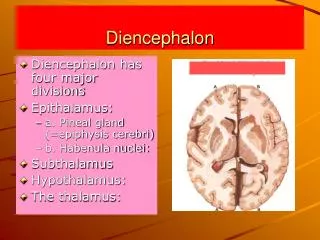
Diencephalon
Diencephalon. Hypothalamus. Objectives. 1.Describe the structure of hypothalamus, epithalamus , and subthalamus 2.List the nuclei of the hypothalamus 3.List the afferent connections of the hypothalamus 4.List the efferent connections of the hypothalamus

Diencephalon
E N D
Presentation Transcript
Diencephalon Hypothalamus
Objectives 1.Describe the structure of hypothalamus, epithalamus , and subthalamus 2.List the nuclei of the hypothalamus 3.List the afferent connections of the hypothalamus 4.List the efferent connections of the hypothalamus 5.List the functions of hypothalamus
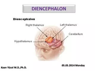
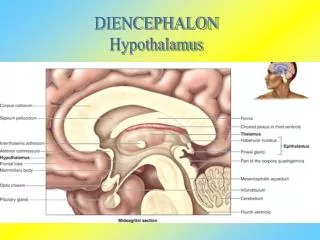
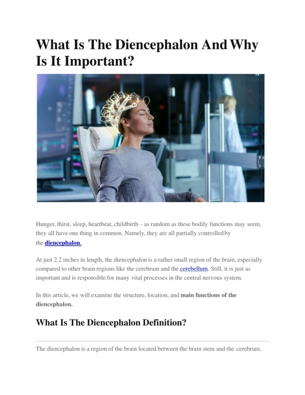
Hypothalamus The hypothalamus lies in the anterior portion of the diencephalon, below the thalamus and above the pituitary gland. It forms part of the wall and floor of the third ventricle. The hypothalamus consists of only 4 cm3 of neural tissue, or 0.3% of the total brain
Hypothalamus Its functional significance is disproportionate to its size. It has been considered as the head nucleus of the ANS as it is the principle autonomic center in the brain. .
Most of the hypothalamus is hidden except the inferior surface, that can be seen on the inferior surface of the brain, cranial to the cerebral peduncles Parts of hypothalamus seen on the base of the brain include: Preoptic area Infundibulum Tuber cinerium Mammillary bodies oc tc I mb P
Subdivisions of the hypothalamus Divided into three zones Periventricular zone Intermediate zone Lateral zone The periventricular and intermediate zones are often described together as medial zone
Subdivisions of the hypothalamus • From medial to lateral: 1-Periventricular zone 2- Intermediate zone 2-Lateral zone Lines the walls of 3rd ventricle, above the pituitary. Divided into medial and lateral regions by the fornix.
The anterior column of the fornix passes vertically through the substance of hypothalamus (to terminate in the mamillary body) and divides it into medial and lateralzones
Subdivisions of the hypothalamus • Anterior to posterior: 1- Preoptic region: Adjoins lamina terminalis which isalayer of gray matter in the brain connecting the optic chiasma and the anterior commissure 2- Suraoptic region: Lies above optic chiasma 3-Tuberal region: (infundibulotuberal) • -includes infundibulum • tubercinereum 4- Mamillary region: consists of mamillary body and area above it
Medial Nuclei Lateral Nuclei
Lateral part • Lies medial and ventral to the subthalamus • Traversed by many fibers including medial forebrain bundle (connecting the hypothalamus with the midbrain tegmentum and the limbic system) • Controls food and water intake (feeding centre) • Lesions cause aphagiaand adipsia
Medial part • Forms lateral wall of the 3rd ventricle • Contains: • Anterior nucleus • Supraoptic nucleus • Preoptic nucleus • Paraventricular nucleus • Dorsomedial nucleus • Ventromedial nucleus • Posterior nucleus • Mammillary nuclei • Infundibularnucleus (Arcuate)
Supraoptic nucleusproduces vasopressin which increases water reabsorption by the kidney Paraventricular nucleusproduces oxytocin The axons of cells in supraoptic and paraventricular nuclei pass to the neurohypophysis in the hypothalamo-hypophyseal tract The hormones are transported in this tract and released into thecapillary bed
Suprachiasmatic nucleus: concerned with the control of diurnal rhythm and sleep/awake cycle Receives some afferent fibers directly from the retina Ventromedial nucleus: acts as satiety centre Lesions cause polyphagia, polydipsia Bilateral lesion of the medial part of the ventromedial nucleus causes hyperphagia and obesity . Further lesion of the lateral part of the ventromedial nucleus in the same person produces complete cessation of food intake.
Mammillary nuclei: Part of the limbic system Receive afferents from the hippocampusthrough fornix Project to the: • Anterior nucleus of thalamus through mammillo-thalamic tract • Brainstem through the mamillotegmental tract
Optic tract Mamillary body Column of fornix Thalamus Superior & inferior colliculi Caudate nucleus Anterior commissure Mamillothalamic tract
Hypothalamus also synthesizes releasing factors & release-inhibiting factors, that control the release of hormones by the adenohypophysis These factors are released from the terminals of hypothalamic neurones into the capillary bed of the pituitary portal system, which conveys the release agents to the anterior pituitary
Functions • Co-ordination of homeostatic mechanism • Controls the release of hormones from the pituitary gland. • Center for regulation of autonomic activity --- controls medulla oblongata nuclei for cardiovascular, respiration • Activation of posterior region associated with sympatheticresponses • Activation of anterior region associated with parasympathetic responses • The mammillary nuclei are associated with the emotional behaviour and memory • The suprachiasmatic nucleus is concerned with diurnal rhythm & sleep/waking cycle • The lateral hypothalamus & the ventromedial nucleus regulate feeding and drinking • Center for Feeding reflexes—licking, swallowing, etc. • Controls subconscious skeletal muscle movements—facial expressions, sexual movements • Coordinates autonomic response to conscious input—thought of fear produces accelerated heart rate, etc.
Connections of the hypothalamus -Hypothalamus is concerned with visceral function -Connected to various parts of limbic system, reticular formation, autonomic centres in brainstem and spinal cord.
Afferent connections The hypothalamus recievesvisceral (including Taste) through spinal cord and brainstem. Afferents from nucleus of tractussolitariusto hypothalamus carry taste sensation. Somatic afferents reach through collaterals of major ascending tracts Afferents from olfactory pathway and limbic system. Anterior perforated substance, septal nuclei Amygdaloid complex, hippocampus, piriform cortex. Neocortex Thalamus Limbic system Hypothalamus Visual input Ascending Somatosensory pathway Visceral centres In brainstem & Spinal cord.
Efferent connections • The hypothalamus sends fibres to autonomic centresin brain and spinal cord • In brainstem:-Nucleus of solitary tract -Dorsal nucleus of vagus -Nucleus ambigus -Parabrachial nucleus
Epithalamus • Relatively small part, located in most caudal and dorsal region • Lies immediately rostral to superior colliculus • Consists of: • Pineal gland & • Habenular nuclei
Pineal Gland • An endocrine organ • Synthesizes melatonin • Controls: • Sleep/awake cycle • Regulation of onset of puberty
Habenular nuclei • Located in habenular triangle (area in the posterior part of the diencephalon, just anterior to pineal gland) • Have connections with limbic system • Serves autonomic function and emotional drives
SUBTHALAMUS • Region of diencephalon located below the thalamus & dorsolateral to hypothalamus • Continues caudally with the midbrain Th Hypothalamus
Contents • Rostral extension of: • Red nucleus • Substantianigra • Brainstem reticular formation asZonaincerta • Long tracts passing through brain stem and heading toward thalamus • Spinothalamic & Trigeminothalamic tracts • Medial lemniscus • Dentatothalamic fibers • Pallidothalamic fibers (fasciculus lenticularis, Ansalenticularis & thalamic fascicle) • Subthalamic nucleus
Subthalamic Nucleus • Resembles a biconvex lens in shape • Located in the ventrolateral part of the subthalamus • Lies against the medial surface of the internal capsule IC
Connections • Has reciprocal connections with ipsilateral: • Globuspallidusvia subthalamic fasciculus, which passes through the internal capsule • Substantianigra
Functions Lesions Rare Usually of cerebrovascular origin Results in Hemiballism(sudden, forceful involuntary, violent or jerky, movements of the limbs) on the contralateral side Plays an important role in normal functioning of basal ganglia
ZonaIncerta • Rostral extension of the brainstem reticular formation • Enveloped by pallidothalamic fibers (lies between the lenticular fascicle and the thalamic fascicle)