Download
1 / 34
430 likes | 800 Vues
Internal Fracture Fixation in Patient with Osteoporosis. Presenter: dr. Nicko Perdana Moderator: Dr. dr. Ismail, SpOT. Osteoporosis . A systemic disease. Primary or secondary

E N D
Internal Fracture Fixation in Patient with Osteoporosis Presenter: dr. Nicko Perdana Moderator: Dr. dr. Ismail, SpOT
Osteoporosis • A systemic disease. • Primary or secondary • Primary occurs in an individual who has no endocrinopathy or other disease state that would account for the changes in bone mass. • Characterized by: • Decreased bone mass. • Deteriorated bone microarchitecture. • In the elderly (≥ 65 years): the most contributing factor (75%) fractures caused by low energy fall.* *Lucas TS, Einhorn TA: Osteoporosis: The role of the orthopaedist. J Am Acad Orthop Surg 1993;1:48-56
Site of Fracture • Generally involves the metaphyseal region of skeleton. • Why metaphyseal? • Composed mostly of cancellous bone. • Greater surface for bone turnover rate (compared with cortical bone). • Proximal femur, distal femur, proximal tibia, distal radius, proximal humerus.
Principles of Fx Management • The goal of definitive fracture carein elderly patients:early restoration of function. • Timely treatment (best condition < 48 hr).* • Evaluation of concurrent illness. • Preoperative management to optimize the condition. • Procedure should be simple & minimal. • Early weight bearing. *Aharonoff GB, Koval KJ, Skovron ML, Zuckerman JD: Hip fractures in theelderly:Predictors of one year mortality. J Orthop Trauma 1997;11:162-165
Principles of Fx Management • The principles of biologic fracture repair should be applied whenever possible: • Careful handling of the soft tissue. • Avoiding unnecessary stripping. • Minimizing exposure of fracture site. • Preservation of fracture hematoma.
Principles of Fx Management • Decline in capacity of fracture repair is age related.* • Disturbance of the development of strength within fracture callus: *Silver JJ, Einhorn TA: Osteoporosis and aging: Current update. Clin Orthop 1995;316:10-20
Problems in Fx Management • Bone failureNOTimplant breakage. • Bone mineral density correlates with holding power of screws. • Osteoporotic bone lacks the strength to hold screw / plate securely. * • Loosening of the screw & implant. * Sjostedt A, Zetterberg C, Hansson T, Hult E, Ekstrom L: Bone mineral content andfixation strength of femoralneck fractures: A cadaver study. Acta Orthop Scand 1994;65:161-165
Solution for Fx Management • Traditional internal fixation techniques must be modified. • IF devices that allow load sharing is used to minimize stress at the bone-implant interface. • Sliding nail plate devices, • intramedullary nails, • antiglides plates, and • tension band constructs BETTER than more rigid techniques.
Implant Fixation • Screws • Plates • Intramedullary nails • Tension band wiring • Augmentation
Screws Resistance to pullout depends on: • Length of the screw • Thread diameter • Quality of the bone • Density • Trabecular orientation • Direction of insertion* • Parallel BETTER than perpendicular to the trabecular pattern. *An YH, Young FA, Kang Q, WilliamsKR: Effects of cancellous bone structureon screw pullout strength. MedicalUniversity of South Carolina Orthopedic Journal 2000;3:22-26
Screws • Bone quality prime determinant of screw holding. • When bone mineral content falls below 0.4 gram/cm2, the effect of varying thread diameter is lost.* • To prevent loosening: • Place it as parralel as possible. • Use the largest thread diameter compatible with the fracture scale. *Turner IG, Rice GN: Comparison ofbone screw holding strength in healthybovine and osteoporotic human cancellous bone. Clin Mater 1992;9:105-107
Screws • In cases of severe osteoporosis screw fixation may be augmented with Polymethylmethacrylate (PMMA).* • (1)Once the cement components are mixed (2)injected into the stripped screw holes (3)place the screw but incompletely tightened (4)after the cement has set (5)the screw is fully tightened. *Motzkin NE, Chao EYS, An K-N, Wikenheiser MA, Lewallen DG: Pullout strength of screws from polymethylmethacrylate cement. J Bone Joint Surg Br 1994;76:320-323
Plates • The strength of plate fixation is affected by the degree of comminution & the resulting size of any gap at fracture site. • The most important factor reducing strain in plated fracture is cortical contact. • Screw spacing is more important than the number of screws used for fixation.* *Törnkvist H, Hearn TC, Schatzker J: The strength of plate fixation in relation to the number and spacing of bone screws. J Orthop Trauma 1996;10:204-208
Plates • Ellis et al * concluded: three screws should be placed adjacent in either side of fracture gap. • Cortical contact at the fracture site is paramount. • In certain conditions (e.g. moderate comminution) the fracture should be shortened to achieve contact. ** * Ellis T, Bourgeault CA, Kyle RF: Screw position affects dynamic compression plate strain in an in vitro fracture model. J Orthop Trauma 2001;15:333-337 ** Blatter G, König H, Janssen M, Magerl F: Primary femoral shortening osteosynthesis in the management of comminuted supracondylar femoral fractures. Arch Orthop Trauma Surg 1994;113:134-137
Plates • Plates act as tension band NOT as bridge. • When comminution is extensive consider double-plating. • In oblique or spiral plates act as antiglide.
Intramedullary nails • Treatment of choice for diaphyseal fractures in osteoporotic bone (femur & tibia). * • The advantages of nailing: ** • Providing broad area of purchase. • Allowing load sharing. • Sufficiently secure fixation to allow immediate weight bearing. * McConnell T, Court-Brown C, Sarmiento A: Isolated tibial shaft fracture. J Orthop Trauma 2000;14:306-308 ** Rodriguez Alvarez J, Casteleiro Gonzolez R, Laguna Aranda R, Ferrer Blanco M, Cuervo Dehesa M: Indications for use of the long Gamma nail. Clin Orthop 1998;350:62-66
Intramedullary nails • IM positioned closer to mechanical axis smaller bending forces than plate (external surface). • Less fatigue failure compared with plate. • Greater strength in axial loading but less stable during bending & torsion. * • Better suited for fixation of severely comminuted osteoporotic bone fracture. * Ito K, Grass R, Zwipp H: Internal fixation of supracondylar femoral fractures:Comparative biomechanical performance of the 95-degree blade plate and two retrograde nails. J Orthop Trauma 1998;12:259-266
Intramedullary nails • Major weakness of interlocking medullary nails loosening of locking screws. • It is likely in distal femur rotational fragment varus / valgus deformity. • Locking screw fixation can be improved: • By using different planes of screw orientation (anteroposterior & transverse placement). • By using cement.
Tension band wiring • TBW is usually applied to transverse fractures, which are distracted. • It provides strong & secure fixation early mobilization of involved joint. • Fractures: patella, olecranon, medial malleolus, proximal humerus.
Augmentation • Bone grafting • Autograft: iliac crest (most common). • Allograft: allograft bone, demineralized allograft bone, synthetic osteoconductive materials. * • Bone cement • PMMA (polymethylmetacrylate) • Calcium phosphate. * Gazdag AR, Lane JM, Glaser D, Forster RA: Alternatives to autogenous bone graft: Efficacy and indications. J Am Acad Orthop Surg 1995;3:1-8
Augmentation Bone autograft (using cancellous bone) • (+) • Cancellous bone can encourage rapid fracture healing osteoinductive, osteoconductive, osteogenic. 30 • Osteoporotic bone is NOT an inferior graft material. • (-) • There is morbidity associated with the harvest of autogenous bone. • Requiring larger exposure to get more bone graft.
Bone Cement • PMAA (Polymethylmethacrylate) • Replacement of severely comminuted areas. • Successfully used in femur supracondylar fx & intertrochanteric fx. • But, it is NOT ideal material permanent, foreign body, generating heat. • Can be used to augment screw fixation. • Calcium phosphate • Adhere better to bone. • Capable of being resorbed & replaced by host bone. • Successfully used in intertrochanteric fx & distal radius fx. • Used to fill the voids, not for augmentation of screw fixation.
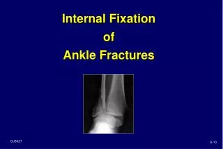
Fracture Types • Intertrochanteric fractures • Supracondyler fractures of the distal femur • Lateral tibial plateu fractures • Ankle fractures & the distal fibula • Proximal humerus fractures
Postoperative Care • Physical rehabilitation & psychososial treatment. • Preinjury functional compromise + additional disability associated with recovery + depression/hopelessness multidisciplinary service. • Malnourished state Clinical evaluation of nutritional status. • Any patient past middle age with low-energy metaphyseal fx: • Undergo BMD testing • Placed on regiment to combat further bone loss. • Calcium 1000-1500 mg/day • Vitamin D • Biphosphonate therapy