Download
1 / 46
510 likes | 878 Vues
Chronic Kidney Disease. Dr Dana Ahmed Sharif MRCP UK/ MRCP London. Each kidney has: a renal artery a renal vein a ureter which enter the kidney at the hilum. The kidney has: an outer cortex layer an inner medullary pyramid layer fine branches of the ureter which form the calyx.

E N D
Chronic Kidney Disease Dr Dana Ahmed Sharif MRCP UK/ MRCP London
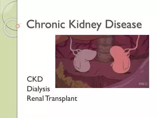
Each kidney has: • a renal artery • a renal vein • a ureter • which enter the kidney at the hilum. • The kidney has: • an outer cortex layer • an inner medullary pyramid layer • fine branches of the ureter which form the calyx
The NEPHRON is the functional unit of the kidney. Each kidney has nearly a million of nephrons Each nephron: is supplied with an afferent arteriole. produces urine which drains into a collecting duct. The collecting ducts fuse together to make larger and larger tubes until they join together as the ureter Each nephron consists of: a glomerulus & Bowman’s capsule a proximal convoluted tubule a loop of Henle a distal convoluted tubule
Chronic Kidney Disease • Longstanding and irreversible decline in renal function • Injured nephrons replaced by extensive fibrosis • Normal nephrons gradually fail due to ‘hyperfilteration’ • Is mainly TUBULAR scarring, so urine output usually normal, though urine quality reduced • Any cause of AKI can eventually lead to CKD
Assessment of renal function • 60 years old woman - Creatinine 1.1 (0.4-1.1mg/dl) - Wt 48 Kg • 20 years old man - Creatinine 1.3 (0.7-1.2mg/dl) - Wt 98 Kg
How do we assess renal function? • Serum urea and creatinine - Small molecules - Filtered by glomerulus – measure of GFR - But creatinine also depends on: + Muscle mass + Body size + Sex and race • Measurement of GFR - Direct measurement - Estimation of GFR (eGFR)
Shenmesh, O., Golbetz, H., Kriss, J. P., and Myers, B. D. (1985). Kidney International 28 , 830–838
Staging of chronic kidney diseaseK/DOQI guideline *Only considered to be CKD stage 1 or 2 if proteinuria and/or haematuria or structural renal disease are present
CKD prevalence Stage 5: 0.2% Stage 4: 0.2% Stage 3: 4.3% Stage 2: 3.0%% Stage 1: 3.3% Coresh J. Astor BC, Greene T, et al. (2003) Prevalence of chronic kidney disease and decreased kidney function in the adult US population: Third National Health and Nutrition Examination Study. Am J Kidney Dis, 41(1), 1-12
Why is it important to identify patients with CKD? • CKD predispose to increased cardiovascular risk (modify risk factors such as Hypertension) • Some patient may benefit from other investigations ( such as renal biopsy) • It may be possible to slow down the progression • Complications of CKD can be identified and treated early • Preparation for dialysis or transplantation once they reach end-stage.
?Acute or Chronic kidney impairment • History and duration of symptoms • Previous urinalysis or measurement of renal function • Rapid changes in renal function • Normocytic, normochromic anaemia( but haemorrhage, haemolysis) • Ultrasound: small kidneys and increased echogenicity • Evidence of renal osteodystrophy ( ie digital subperiosteal erosions)
Who is at risk? • Known CKD • Hypertension • Diabetes • Unexplained oedema • Congestive cardiac failure • Atherosclerotic disease ( coronary, cerebral, peripheral) • Multi systemic disease ( myeloma, SLE) • Bladder outflow obstruction, neurogenic bladder, renal stone disease, recurrent UTI • Chronic nephrotoxic use ( NSAID, ACEI, ARBII, ciclosporin, lithium) • Urologically unexplained haematuria
Why chronic renal failure tends to progress? • Once CKD established, inexorable progression is likely • While only 10% of those with CKD stage 3 may progress, all are at risk • Therefore life-long follow up is desirable
Mechanism of progression • Raisedintraglomerular pressure • Glomerular damage • Proteinuria • Tubulo-intestitial scarring
Importance of proteinuria • Normal glomeruli do not leak protein • Proteinuria is a sign of glomerular dysfunction • Proteinuria is also risk factor for progression of CKD
Importance of proteinuria • Normal glomeruli do not leak protein • Proteinuria is a sign of glomerular dysfunction • Proteinuria is also risk factor for progression of CKD • Measures which reduce proteinuria slow down the progression of CKD* • Albuminuria is a marker of cardiovascualr risk** *GISEN Group (1997). Lancet 349 , 1857–1863 **Hebert, L. A., Spetie, D. N., and Keane, W. F. (2002). Postgraduate Medicine 111 , 23.
Causes of End Stage Renal Disease* • Diabetes • Glomerulonephritis • Reflux nephropathy • Renovascular disease • Hypertension • Polycystic kidney disease *UK renal data registry
Case 1 • 55 years old male • History of diabetes for 15 years • He has CKD with eGFR 35ml/min( checked 6 months ago). • His BP was 140/90 and his doctor started him on Ramipril 2.5 mg two weeks ago. • Lab results: eGFR of 25ml/min otherwise well, HbA1c 6.0%, urinary protein excretion of 0.5g/24hrs( no changes from previous results).
What is the most likely cause of his renal deterioration? 1- Progression of his diabetic nephropathy 2- Uncontrolled hypertension 3- Infection 4- Drug induced
What is the most likely cause of his renal deterioration? 1- Progression of his diabetic nephropathy 2- Uncontrolled hypertension 3- Infection 4- Drug induced
Reversible causes of deteriorating renal function • Blood pressure control • Diabetes control • Obstruction • Drug, contrast agent • Intercurrent illness • Exacerbation of underlying kidney disease (sarcoidosis, lupus)
Preventing progression • Blood pressure and diabetes - Uncontrolled Hypertension and diabetes makes proteinuria worse - Controlling hypertension and diabetes is vital • ACE inhibitors and ARBs have anti-proteinuric effects over and above their effects via blood pressure - Agents of choice in proteinuric renal disease - Even if blood pressure ‘normal’
Blood pressure targets* • CKD with no proteinuria: <140/90 mmHg • CKD with proteinuria: <130/80 mmHg • Diabetic nephropathy: <130/80mmHg (aim for 125/75 mmHg). * NICE guideline for management of Chronic Kidney Disease, Sept 2008
Managing CKD 1- General advice: • Smoking cessation • Weight reduction if obese • Encourage aerobic exercise • Aspirin 75mg od ( if 10 years cardiovascular risk>20%) • Treat hyperlipidaemia • Avoid NSAID and other nephrotoxic drugs • Limit alcohol intake( <21units/wk in male, <14units/wk in female • Vaccination against influenza and pneumococcus
Managing CKD 2- CKD stages 1-3: • Monitor eGFR, urinalysis and protein creatinine ratio • Meticulous BP and diabetes control • If Hb < 11g/dl check ferritin, B12 and folate( if ferritin <100mg/dl start on oral or IV iron) • Annual check of PTH, Calcium and phosphate
Managing CKD 3- CKD stages 4-5: • Refer to nephrologist • Full dietary assessment • Optimize PTH, Calcium and phosphate • Correct acidosis • Hepatitis B immunization • Discuss future treatment (dialysis, transplant or conservative/palliative)
Complications of CKD • Uraemia • Anaemia • Renal bone disease • Salt and water retention • Electrolyte imbalance • Acidosis • Cardiovascular disease and lipids
Salt and water retention • Dietary salt restriction ( aim <5g/day) • Fluid intake restriction • Diuretics such as furosemide, titrate up • Add thiazide ( metolazone) • Those with fluid overload and on diuretics monitor: - Weight ( loss of 1kg/day) - BP control - Monitor electrolyte - if Ur > 70mg/dl consider reducing diuretics - Refractory volume overload = dialysis
Hyperkalaemia • Common and potentially fatal • Rapid rise of K+ is more dangerous than gradual ones, as cell membrane stability is more vulnerable to acute changes • ↑K+ → depolarization of the membrane resting potential → Na+ channel inactivation → ↓membrane excitability → neuromuscular depression and cardiac dysrhythmias. • When to treat: • K+: 5.5-6.0 mmol/l: recheck routinely. Review medications and arrange dietary advice • K+: 6.1-6.5 mmol/l: recheck urgently, review med, stop ACEI/ARB2 and arrange dietary advice. • K+: >6.5 mmol/l: admit for emergency management
Measures to prevent ↑K+ - Dietary restriction: diary products, potatoes and some fruits (such as banana ,grapes, pineapple), fresh fruit juice, tomatoes, sweet corn, mushrooms, chocolate and coffee. - Loop diuretics: promote urinary K+ excretion. - Drug withdrawal or dose reduction (ACEI, ARB2), review other contributory drugs (spironolactone, NSAID and B- blockers) - Correct acidosis. - Dialysis ( especially in refractory ↑K+)
Acidosis Leads to: • Bone: increase bone resorption and impaired mineralization, contributing to renal osteodystrophy. • Over ventilation (compensatory mechanism) • Hyperkalaemia • Increased ionized Calcium (acidosis lead to reduced albumin bound fraction) • Malnutrition: acidosis promotes catabolism
How to correct acidosis? • Treat when venous HCO3- is <21 mmol/l • Give NaHCO3 tablet 0.5-1.5g/tds • Refractory acidosis: dialysis
Uraemia • Clinical syndrome caused by substantial fall in GFR • Failure to eliminate potentially toxic small and middle size molecules. • NOT a result of high blood urea concentration. • Ur and Cr are not directly toxic. • Leads to: - Chronic inflammation and ↑oxidative stress - Accumulation of metabolic end products - Accelerated atherogenesis - Disruption of immune system - phosphate is retained: leads to ↑PTH, arteriosclerosis and vascular calcification
Uraemia Symptoms: • Nausea/ vomiting • Anorexia and weight loss • Malaise, fatigue • Confusion, fits and coma • Pericarditis
Anaemia • Erythropoietin(EPO) is essential for the terminal maturation of erythrocyte • EPO deficiency occur in most advanced CKD ( eGFR < 35ml/min) with exception: - Adult polycystic kidney disease - Benign renal cysts - Renal cell carcinoma
Differential diagnosis of anaemia in CKD • EPO deficiency • Iron deficiency anaemia • Blood loss ( GI tract, Haemodialysis) • Folate deficiency • B12 deficiency • Haemolysis • Myelodyplasia • Myeloma
Case 2 • 54 years old male with CKD stage 4 • History or recurrent chest infection for the last 3months • On oral co-amoxiclav, Erythropoietin injection 6000 iu sc weekly( increased recently) • Lab results: Hb 8.5g/dl(no changes), WBC: 14, eGFR:16, CRP:75, ferritin: 600
What is the most likely cause of EPO non-responsiveness 1- Non-compliance 2- Inadequate dose of EPO 3- High CRP 4- Iron deficiency anaemia
What is the most likely cause of EPO non-responsiveness 1- Non-compliance 2- Inadequate dose of EPO 3- High CRP 4- Iron deficiency anaemia
EPO non-response • Iron deficiency • Chronic blood loss • Infection/inflammation • Severe hyperparathyroidism (causes bone marrow fibrosis) • B12 and folate deficiency • Haemolysis • Malnutrition • Inadequate dosing • Poor compliance
Renal bone disease (Osteodystrophy) • Heterogeneous disorder leading to diminished bone strength • Is a function of bone turnover, density, mineralization and architecture • Mostly occur beyond CKD stage 3 • High turnover( PTH > 450ng/L*) : caused by secondary hyperparathyroidism leads to increased bone resorption and formation(osteitisfibrosacystica) • Low turnover (Adynamic bone disease)( PTH < 100ng/L): paucity of cells with decreased bone resorption and formation. • Osteomalacia: defect in mineralization, deficiency of 1,25(OH)2D • Osteoporosis: reduced bone density
Renal bone disease: clinical features 1- Secondary hyperparathyroidism: • Usually asymptomatic • Bone pain and arthralgia • Muscle weakness • Pruritis (cutaneous calcium phosphate deposition) • Bone deformity • Increased fracture risks (hip fracture x5 in Dialysis) • Marrow fibrosis contribute to anaemia 2- Adynamic bone disorder: asymptomatic and may have twice increase in fracture risk than dynamic bone disease. 3- Increased cardiovascular risk
Renal bone disease: treatment • Measures to reduce serum phosphate: - Dietary PO4 restriction( meat, egg, milk, cereals) - Dialysis - Oral phosphate binders( prevent absorption)such as Ca+2 and non-Ca+2 containing phosphate binder • Measures to increase serum calcium and suppress PTH: - Ca+2 salts ( also act as PO4 binder) - Vit D analogues (calciterol, alfacalcidol) • Measures to suppress PTH directly: - Calcimimetic agents(cinacalcet) - Parathyroidectomy( tertiary PTH= PTH, Ca2,PO4 and ALP)
Malnutrition Causes: • Anorexic uraemic toxins(↑leptin) • Chronic low grade inflammation and oxidative stress. • Dietary restriction (low protein diet) • Medications (iron, PO4 binders) • Acidosis • Dialysis itself (protein loss during dialysis) • Biochemical indicators: s. albumin, transferrin and cholesterol