Viruses
530 likes | 649 Vues
This text provides a comprehensive overview of viruses, detailing their structure, classification, and how they multiply within host cells. Viruses consist of DNA or RNA enclosed in a protein coat, and some have an additional envelope. The classification of viruses is based on their morphological types, with taxonomy that includes various family and genus names. Additionally, the text explores the different multiplication cycles of bacteriophages and animal viruses, illustrating the processes of attachment, penetration, biosynthesis, maturation, and release, using examples such as retroviruses and herpesviruses.

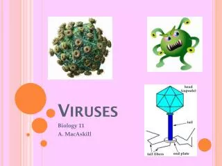
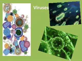
Viruses
E N D
Presentation Transcript
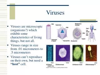
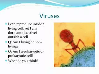
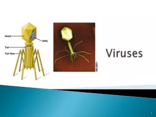
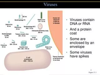
Viruses • Viruses contain DNA or RNA • And a protein coat • Some are enclosed by an envelope • Some viruses have spikes Figure 13.1
Viruses • Host range is the spectrum of host cells that a virus can infect. • Most viruses infect only specific types of cells in one host • Host range is determined by specific host attachment sites and cellular factors
Viral Structure • Virion is a fully developed, infectious viral particle composed of • Capsid or protein coat • Nucleic acid core • Envelope (not present on all viruses) • The arrangement capsid can be used to classify viruses into several different morphological types.
Helical Viruses Figure 13.4a, b
Polyhedral Viruses Figure 13.2a, b
Complex Viruses Figure 13.5a
Viral Taxonomy • Family names end in -viridae • Genus names end in -virus • Order names end in –ales • Viral species: A group of viruses sharing the same genetic information and ecological niche (host). • Common names are used for species • Subspecies are designated by a number
Viral Taxonomy • Herpesviridae • Herpesvirus • Human herpes virus 1, HHV 2, HHV 3 • Retroviridae • Lentivirus • Human Immunodeficiency Virus 1, HIV 2
Growing Bacterial Viruses • Viruses must be grown in living cells. • Plaque Assay is used to quantitate virus. • Bacteriophages form plaques when grown on agar. • Plaques are cleared areas in the bacterial lawn. Figure 13.6
Growing Animal Viruses • Animal viruses may be grown in: • Living animals • Embryonated eggs. Figure 13.7
Growing Animal Viruses • Animal and plants viruses may be grown in cell culture. • Primary Cell lines tend to die out after a few generations. • Continuous cell lines may be maintained indefinitely. Figure 13.8
Virus Identification • Cytopathic effects • Serological tests • Detect antibodies against viruses in a patient • Use antibodies to identify viruses in neutralization tests, viral hemagglutination, and Western blot • Nucleic acids • RFLPs • PCR
Virus Identification Figure 13.9
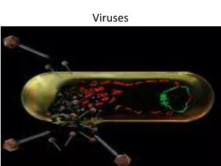
Multiplication of Bacteriophages • Lytic cycle Phage causes lysis and death of host cell • Lysogenic cycle Prophage DNA incorporated in host DNA
Multiplication of Bacteriophages (Lytic Cycle) • Attachment Phage attaches by tail fibers to host cell • Penetration Phage lysozyme opens cell wall, tail sheath contracts to force tail core and DNA into cell • Biosynthesis Production of phage DNA and proteins • Maturation Assembly of phage particles • Release Phage lysozyme breaks cell wall
Multiplication of Bacteriophages (Lytic Cycle) Bacterial cell wall Bacterial chromosome Capsid DNA Capsid Sheath Tail fiber Tail 1 Attachment:Phage attaches to host cell. Base plate Pin Cell wall Plasma membrane 2 Penetration:Phage pnetrates host cell and injects its DNA. Sheath contracted Tail core 3 Merozoites released into bloodsteam from liver may infect new red blood cells Figure 13.10.1
Multiplication of Bacteriophages (Lytic Cycle) Tail DNA 4 Maturation:Viral components are assembled into virions. Capsid 5 Release:Host cell lyses and new virions are released. Tail fibers Figure 13.10.2
Viral Growth Curve Figure 13.11
The Lysogenic Cycle Figure 13.12
Specialized Transduction gal gene Bacterial DNA Prophage 1 Prophage exists in galactose-using host (containing the gal gene). Galactose-positive donor cell gal gene 2 Phage genome excises, carrying with it the adjacent gal gene from the host. 3 Phage matures and cell lyses, releasing phage carrying gal gene. gal gene 4 Phage infects a cell that cannot utilize galactose (lacking gal gene). Galactose-negative recipient cell 5 Along with the prophage, the bacterial gal gene becomes integrated into the new host’s DNA. 6 Lysogenic cell can now metabolize galactose. Galactose-positive recombinant cell Figure 13.13
Multiplication of Animal viruses • Attachment Viruses attaches to cell membrane • Penetration By endocytosis or fusion • Uncoating By viral or host enzymes • Biosynthesis Production of nucleic acid and proteins • Maturation Nucleic acid and capsid proteins assemble • Release By budding (enveloped viruses) or rupture
Attachment, Penetration, and Uncoating Figure 13.14
Release of an enveloped virus by budding Figure 13.20
Multiplication of DNA Virus Papovavirus 1 Virion attaches to host cell 7 Virions are released Host cell DNA Capsid 2 DNA Virion penetrates cell and its DNA is uncoated Cytoplasm 6 Virions mature Capsid proteins mRNA 5 Late translation; capsid proteins are synthesized 3 Early transcription and translation; enzymes are synthesized 4 Late transcription; DNA is replicated Figure 13.15
Pathways of Multiplication for RNA-Containing Viruses Figure 13.17
Multiplication of a Retrovirus Capsid Reverse transcriptase DNA Virus Two identical + stands of RNA 1 Retrovirus penetrates host cell. Host cell DNA of one of the host cell’s chromosomes 5 Mature retrovirus leaves host cell, acquiring an envelope as it buds out. Reverse transcriptase 2 Virion penetrates cell and its DNA is uncoated Viral RNA Identical strands of RNA 4 Transcription of the provirus may also occur, producing RNA for new retrovirus genomes and RNA that codes for the retrovirus capsid and envelope proteins. Viral proteins RNA 3 The new viral DNA is tranported into the host cell’s nucleus and integrated as a provirus. The provirus may divide indefinitely with the host cell DNA. Provirus Figure 13.19
Viruses and Cancer • Viruses have been shown to cause cancer. • Viruses integrate into host cell DNA, causing DNA damage. • Oncogenes are alterations to normal cellular DNA that lead to cancer development
Cancer • Activated oncogenes transform normal cells into cancerous cells. • Oncogenes can be activated by a variety of agents. • Transformed cells have increased growth, loss of contact inhibition, tumor specific transplant and T antigens.
Oncogenic Viruses • Oncogenic RNA viruses • Retroviridae • Viral RNA is transcribed to DNA which can integrate into host DNA • HTLV 1 • HTLV 2 • Oncogenic DNA Viruses • Adenoviridae • Heresviridae • Poxviridae • Papovaviridae • Hepadnaviridae
Viral Infections • Latent Viral Infections • Virus remains in asymptomatic host cell for long periods • Cold sores, shingles • Persistent Viral Infections • Disease processes occurs over a long period, generally fatal • Subacute sclerosing panencephalitis (measles virus)
Prions • Infectious proteins • Inherited and transmissible by ingestion, transplant, & surgical instruments • Spongiform encephalopathies: Sheep scrapie, Creutzfeldt-Jakob disease, Gerstmann-Sträussler-Scheinker syndrome, fatal familial insomnia, mad cow disease • PrPC, normal cellular prion protein, on cell surface • PrPSc, scrapie protein, accumulate in brain cells forming plaques
Prions PrPSc PrPc 2 3 4 1 Lysosome Endosome 5 6 7 8 Figure 13.21
Plant Viruses • Plant Viruses • Plant viruses enter through wounds or via insects • Viroids • Viroids are infectious RNA; potato spindle tuber disease Figure 13.22
Virus Families • Single-stranded DNA, nonenveloped viruses • Parvoviridae • Human parvovirus • Fifth disease • Anemia in immunocompromised patients
Double-stranded DNA, nonenveloped viruses • Mastadenovirus • Respiratory infections in humans • Tumors in animals
Double-stranded DNA, nonenveloped viruses • Papillomavirus (human wart virus) • Polyomavirus • Cause tumors, some cause cancer
Double-stranded DNA, nonenveloped viruses • Orthopoxvirus (vaccinia and smallpox viruses) • Molluscipoxvirus • Smallpox, molluscum contagiosum, cowpox
Double-stranded DNA, nonenveloped viruses • Simplexvirus (HHV1 and HHV 2) • Varicellavirus (HHV 3) • Lymphocryptovirus (HHV 4) • Cytomegalovirus (HHV 5) • Roseolovirus (HHV 6) • HHV 7 • Kaposi's sarcoma (HHV 8) • Some herpesviruses can remain latent in host cells
Double-stranded DNA, nonenveloped viruses • Hepadnavirus (Hepatitis B virus) • Use reverse transcriptase to produce DNA from mRNA
Single-stranded RNA, + strand, nonenveloped • Enterovirus • Enteroviruses include poliovirus and coxsackievirus • Rhinovirus • Hepatitis A virus
Single-stranded RNA, + strand, nonenveloped • Hepatitis E virus • Norovirus (Norwalk agent) causes gastroenteritis
Single-stranded RNA, + strand, nonenveloped • Alphavirus • Alphaviruses are transmitted by arthropods; include EEE, WEE • Rubivirus (rubella virus)
Single-stranded RNA, + strand, nonenveloped • Arbovirusescan replicate in arthropods; include yellow fever, dengue, SLE, and West Nile viruses • Hepatitis C virus
Single-stranded RNA, + strand, nonenveloped • Coronavirus • Upper respiratory infections
Single-stranded RNA, – strand, one RNA strand • Vesiculovirus • Lyssavirus (rabies virus) • Cause numerous animal diseases
Single-stranded RNA, – strand, one RNA strand • Filovirus • Enveloped, helical viruses • Ebola and Marburg viruses
Single-stranded RNA, – strand, one RNA strand • Paramyxovirus • Morbillivirus • Paramyxovirus causes parainfluenza, mumps and Newcastle disease
Single-stranded RNA, – strand, one RNA strand • Hepatitis D virus • Depends on coinfection with Hepadnavirus
Single-stranded RNA, – strand, multiple RNA strands • Influenzavirus (Influenza viruses A and B) • Influenza C virus • Envelope spikes can agglutinate RBCs
Single-stranded RNA, – strand, multiple RNA strands • Bunyavirus (CE virus) • Hantavirus