Download
1 / 9
90 likes | 384 Vues
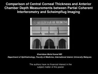
Comparison of Central Corneal Thickness and Anterior Chamber Depth Measurements between Partial Coherent Interferometry and Scheimpflug Imaging. Khairidzan Mohd Kamal MD Department of Ophthalmology, Faculty of Medicine, International Islamic University Malaysia.

E N D
Comparison of Central Corneal Thickness and Anterior Chamber Depth Measurements between Partial Coherent Interferometry and Scheimpflug Imaging Khairidzan Mohd Kamal MD Department of Ophthalmology, Faculty of Medicine, International Islamic University Malaysia The authors have no financial interest in the subject matter of this poster
Introduction Accurate and reliable determination of anterior segment biometry is important in laser and lens refractive surgery Central Corneal Thickness Anterior Chamber Depth • Indicator for corneal metabolism and hydration status • Important parameter in characterization of corneal disease • Basic parameter in the planning of laser refractive surgery • Determine the actual position of the lens. • Assessment of accommodation-induced configuration changes of the phakic eye • Useful parameter in the planning of lens refractive surgery Ultrasound pachymetry has been the traditional “gold standard” for biometry measurement. However, the are newer non-contact optical technologies available. Biometric Technology Slit scanning Anterior Segment OCT Partial Coherent Interferometry Scheimpflug Imaging We sought to evaluate the comparability of different non-contact methods for the analysis of the central corneal thickness and anterior chamber depth in the phakic eye
Purpose Central Corneal Thickness The study is to compare two different non-contact In measuring Partial Coherent Interferometry Short coherence infrared beam is emitted by luminescent diode. Using Michelson interferometer, the beam is separated into two partial rays with different optical paths length These partial rays are reflected onto different intraocular structures. The path difference between the partial rays is recorded when interference signal occurs and smaller than coherence length Anterior Chamber Depth Scheimpflug Imaging Sectional image were measured and recorded CCD camera rotates and provides section planes from three spatial planes The measured data obtained are used to calculate a 3D model from which the thickness and the anterior chamber depth can be computed Methods Strategy Subjects Tools Measurements • Cross sectional • Prospective • 108 healthy eyes • 54 subjects aged 20-25 years old • IIUM Eye Specialist Clinic Kuantan Pahang. • Partial coherent Interferometry • Scheimpflug Imaging • Central Corneal Thickness • Anterior Chamber Depth
Result PCI and SI The mean difference and SD of CCT were -18.63µm+5.76. There was a significant difference in CCT measurement between Partial Coherent Interferometry (PCI) and Scheimpflug imaging (SI), t=-33.59 (107), p<0 .05. PCI SI PCI and SI The mean difference and SD of ACD were 0.059mm+0.249. There was a significant difference in the mean ACD between Partial Coherent Interferometry (PCI) and Scheimpflug imaging (SI), t=2.503 (107), p<0.05. PCI SI
Result PCI PCI SI SI In Pearson’s correlation, there was a linear and positive correlation between Partial Coherent Interferometry (PCI) and Scheimpflug Imaging (SI) in both CCT (p<0.05) and ACD measurement (p<0.05). The observed r in CCT measurement was (0.978) suggests positive and strong correlation. The observed r in ACD measurement was (0.399) suggests positive and fair correlation.
Result In Bland –Altman analysis, a total of 104/108 (96%) Partial Coherent Interferometry-Scheimpflug Imaging differences were within the 95% confidence limits of the mean difference of CCT. A total of 99/108 (92%) Partial Coherent Interferometry-Scheimpflug Imaging differences were within the 95% confidence limits of the mean difference of ACD. SI SI SI PCI PCI PCI PCI SI SI
In short….. • There was overestimation of CCT by Scheimpflug Imaging from Partial Coherent Interferometry and underestimation of ACD by Partial Coherent Interferometry from Scheimpflug Imaging. • The mean difference (95% CI) of CCT was 18.63µm+5.76 with p value <0.05. • The mean difference (95% CI) of ACD was 0.059mm+0.249 with p value=0.014. • Correlation was high to moderate between Partial Coherent Interferometry and Scheimpflug Imaging (r=0.978, r=0.399) with regard to CCT and ACD, respectively.
Discussion • Scheimpflug Imaging (SI) overestimated Partial Coherent Interferometry (PCI) in CCT measurement while PCI overestimated SI in ACD measurement. • The overestimation of CCT measurement between these instruments was statistically and clinically significant. • The estimation of ACD measurement between these instruments however, is statistically significant but not clinically significant. • The comparison between findings in present study and findings in previous studies cannot be made precisely and in quantitatively manner, as different subjects were involved in each study and differences in methodology of study. • The discrepancies between the two instruments could be due to the difference in technological principles, optical measuring technique and operating technique.
References • Buehl, W., Stojanac, D., Sacu, S., Drexler, W., & Findl, O. (2006).Comparison of three methods of measuring corneal thickness and anterior chamber depth. American Journal of Ophthalmology, 141, 1. • Meinhardt, B., Stachs, O., Stave, J., Beck, R., & Guthoff, R. (2006). Evaluation References • of biometric methods for measuring the anterior chamber depth in the non-contact mode. Graefe’s Arch Clinical Experimental Ophthalmology, 244, 559-564. • Wolffsohn, J. S., & Peterson, R. C. (2006). Anterior ophthalmic imaging. Clinical and Experimental Optometry, 89, 4, 205-214. • Wolffsohn, J. S., & Davies, L. N. (2007). Advances in anterior segment imaging. Current Opinion in ophthalmology, 18, 32-38. • Konstantopoulos, A., Hossain, P., & Anderson, D. F. (2007). Recent advances in ophthalmic anterior segment imaging: a new era for ophthalmic diagnosis?. British Journal Ophthalmol, 91, 551-557. • Lackner, B., Schmidinger, G., Skorpik, C. (2005). Validity and repeatability of anterior chamber depth measurements with Pentacam and Orbscan. Optometry and Vision Science, 82, 9, 858-862.