Download
1 / 22
240 likes | 418 Vues
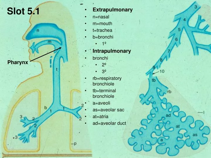
Slot 5.1. Extrapulmonary n=nasal m=mouth t=trachea b=bronchi 1º Intrapulmonary bronchi 2º 3º rb=respiratory bronchiole tb=terminal bronchiole a=aveoli as=aveolar sac at=atria ad=aveolar duct. Slot 1: Diagram of Respiratory System. Pharynx.

E N D
Slot 5.1 • Extrapulmonary • n=nasal • m=mouth • t=trachea • b=bronchi • 1º • Intrapulmonary • bronchi • 2º • 3º • rb=respiratory bronchiole • tb=terminal bronchiole • a=aveoli • as=aveolar sac • at=atria • ad=aveolar duct Slot 1: Diagram of Respiratory System Pharynx
Olfactory epithelia; no goblet cells Thicker than respiratory lining Slot 5.2: Nasal conchae Sustentacular cells Goblet cells Respiratory Epithelium - 1/3 height nuclear free border Basal Cells
Slot 5.3: Larynx Esophagous Epiglottis Stratified Squamous Respiratory Epithelium Cartilage T = trachea Vocalis Muscle Vocal ligament position
Hyaline cartilage Slot 5.4: Trachea • A=adventitia • C=cartilage • P=pericondrial membrane • Green=submucosa • Yellow line = elastic membrane • Pink=respiratory epithelium • E=pseudostratified epithelial cells (has goblet) • L=lamina propria • M=trachealis muscle (smooth) Fibrocartilagenous coat
Slot 5.5: Trachea-bronchi junction • LB=left bronchi • (1º branch) • RB=right bronchi • (1º branch) • C=cartilage • T=trachea
Slot 5.6: Sections of Intrapulmonary bronchus • m=muscularis mucosa • c=cartilage • Arrowhead: mast cells • v=capillaries Submucosa Lamina propria Respiratory epithelium Mucoserous glands
arrows=patches of cilia • arrowheads= clara cells (secret surfactant) • m=muscularis mucosa • a=alveoli • Deep folds do not exist in live animals • c= clara cell Slot 5.7 Bronchioles
Slot 5.8: Lung Diagram Simple cuboidal • Green line = simple cuboidal epithelium; No goblet cells • tb=terminal bronchioles • rb=respiratory bronchioles • A=alveoli, At=atrium, Ad=alveolar duct • Arrows=aveolar pores • s=alveolar sac • (2)=simple squamous
Slot 5.9: Alveoli • A=alveoli p=pulmonary macrophage • e=endothelial cell f=fibroblast cell • s=septa between alveoli g=type II pneumocyte (surfactant) • arrowhead= diffusion barrier
Slot 5.10: Nasal conchae Nerve Bowman’s gland (serous) Microvilli Olfactory cells Basal cells Lamina propria Sustentacular cells Nuclear free border (1/5)
N=nerve • D=duct • B= turbinate bone • V=dilated blood vessels Slot 5.11: Nasal conchae Respiratory epithelium
Slot 5.12: Larynx: sagittal section Epiglottis Vocalis Muscle
submucosa Hyaline cartilage adventitia Lamina propria Slot 5.13 Trachea (c.s.) Respiratory epithelium artifact
Slot 5. 14: Extrapulmonary Bronchus Lamina propria Respiratory epithelium M. mucosa Submucosa Vein Hyaline cartilage
Slot 5.15: Intrapulmonary Bronchus Ciliated Respiratory epithelium Lamina propria Cartilage Muscularis mucosa Alveolar sacs
Slot 5.17: Bronchiole Artery Bronchiole M. mucosa Alveolar ducts and sacs
Slot 5.18: Alveolar duct, alveoli Ad Alveolar pockets Vessel Respiratory bronchiole Simple cuboidal
Slot 5.19: Alveoli Pulmonary macrophage Septa
Slot 5.22: Lung of non-smoker • Note: macrophages have little particulate matter Pulmonary Macrophages
Slot 5.23: Lung of smoker • Note: pulmonary macrophages full of particulate matter (darker staining) Pulmonary macrophages