Download
1 / 72
920 likes | 1.81k Vues
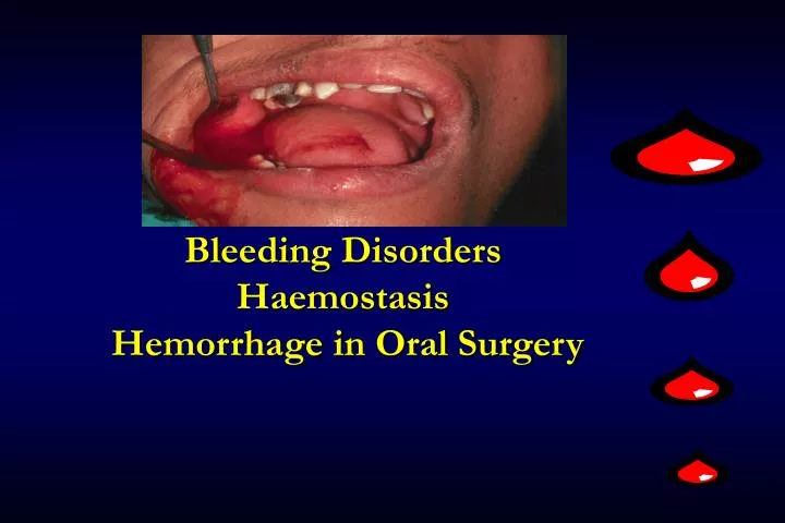
Bleeding Disorders Haemostasis Hemorrhage in Oral Surgery. What is meant by Hemorrhage ?. Prolonged or Uncontrolled Bleeding The amount of blood lost as a result of hemorrhage can range from minimal to significant quantities.

E N D
What is meant by Hemorrhage ? Prolonged or Uncontrolled Bleeding The amount of blood lost as a result of hemorrhage can range from minimal to significant quantities. Hemorrhage can occur to a greater or lesser degree during all surgical procedures and it’s management depends upon whether the patient is hematologically normal or suffers from some disturbance in the normal clotting mechanism.
Hemorrhage in Oral Surgery • The overwhelming majority of patients who undergo oral surgical procedures are those who have normal haemostatic mechanism. • Therefore, significant or major hemorrhages are not that common in oral surgery except in patients who have a bleeding / clotting disorder or those who are on anticoagulants. • However, uncontrolled and persistent bleeding can occur in some healthy patients after dental extraction. • Therefore, it is still important to achieve proper hemostasis in all patients during oral surgical procedures, so as to prevent excessive post-operative blood loss.
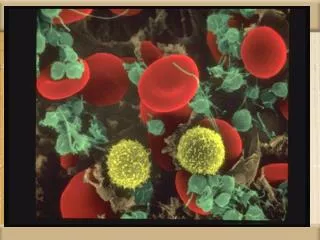
Normal Mechanism of Hemostasis • Hemostasis is a complicated process. • It involves a number of events 1.VASCULAR PHASE 2. PLATELET PHASE 3. COAGULATION PHASE
Hemostasis 1. Blood vessel 2. Blood platelet 3. Blood coagulation 4. Fibrinolysis
Coagulation Cascade Intrinsic system (surface contact) Extrinsic system (tissue damage) XII XIIa Tissue factor XIa XI IX IXa VIIa VII VIII VIIIa X Xa V Va (Thrombin) IIa IIa II Fibrinogen Fibrin Vitamin K dependant factors
Normal Mechanism of Hemostasis • VASCULAR PHASE :When a blood vessel is damaged, Vasoconstriction results. • PLATELET PHASE : Platelets adhere to the damaged surface and form a temporary plug. • COAGULATION PHASE :Through two separate pathways, the Intrinsic and Extrinsic, the conversion of fibrinogen to fibrin is complete. Fibrin tightly binds the platelets to form a clot
HEMOSTASIS DEPENDENT UPON: • Vessel Wall Integrity • Adequate Numbers of Platelets • Proper Functioning Platelets • Adequate Levels of Clotting Factors • Proper Function of Fibrinolytic Pathway
Hemostasis • All component onset at same time and close at different time • Primary(3-5 mins to onset) • Vessel • Platelet • Secondary( 5-10 mins to onset) • Coagulation • Fibrinolysis( need 2-3 days to onset)
Terminology in Bleeding Disorders Petechiae- Pinpoint hemorrhage (Platelets Disorder ) Purpura–Larger, less regular (Platelets Disorder ) Ecchymoses–Over 2 cm – bruise (Coagulation disorders ) Hematoma –Blood trapped in soft tissue (Coagulation disorders ) 10
Petechiae Do not blanch with pressure (cf. angiomas)Not palpable (cf. vasculitis)
Ecchymoses (Typical of Coagulation Factor Disorders)
Specific Symptoms Symptoms Defect of vessel or plt Defect of coag. Organ of frequent bleeding Skin, mucous membrane, muscle, joint nose, gut Size of purpura Small-medium large Bleed at injury sizing Small large Size of bleeding point Petechiae hematoma Ecchimosis Superficial, small deep, large to large Bleeding at Frequent, prolonged Not severe superficial wound Start bleeding Immediately Many hrs after after injured When wound Recurrent bleeding Bleeding stops suppressed when stop suppressing permanently
Clinical Features of Bleeding Disorders Platelet Coagulation disorders factor disorders Site of bleeding Skin Deep in soft tissues Mucous membranes (joints, muscles) (epistaxis, gum, vaginal, GI tract) Petechiae Yes No Ecchymoses (“bruises”) Small, superficial Large, deep Hemarthrosis / muscle bleeding Extremely rare Common Bleeding after cuts & scratches Yes No Bleeding after surgery or trauma Immediate, Delayed (1-2 days), Usually mild Often severe
Inherited bleeding disorders Hemophilia A and B Von Willebrands disease Other factor deficiencies Acquired bleeding disorders Liver disease Vitamin K deficiency/warfarin overdose DIC Coagulation Factor Disorders
Hemophilia A and B Hemophilia A Hemophilia B Coagulation factor deficiency Factor VIII Factor IX Inheritance X-linked X-linked recessive recessive Incidence 1/10,000 males 1/50,000 males Severity Related to factor level <1% - Severe - spontaneous bleeding 1-5% - Moderate - bleeding with mild injury 5-25% - Mild - bleeding with surgery or trauma Complications Soft tissue bleeding
Hemophilia Clinical manifestations (hemophilia A & B are indistinguishable) Hemarthrosis (most common) Fixed joints Soft tissue hematomas (e.g., muscle) Muscle atrophy Shortened tendons Other sites of bleeding Urinary tract CNS, neck (may be life-threatening) Prolonged bleeding after surgery or dental extractions
von Willebrand disease • The most common hereditary bleeding disorder • Often mild and subtle symptoms • Incidence: 1/100, male = female • Autosomal Dominant disorder (12p) • Clinical presentation Epistaxis, ecchymoses, petechiae, menorrhagia, post-operative bleeding
Treatment of von Willebrand disease • Cryoprecipitate • Source of fibrinogen, factor VIII and VWF • Only plasma fraction that consistently contains VWF multimers • Correction of bleeding time is variable • DDAVP (Deamino-8-arginine vasopressin) • Increases plasma VWF levels by stimulating secretion from endothelium • Duration of response is variable • Used for type 1 disease • Dosage 0.3 µg/kg q 12 hr IV • Factor VIII concentrate (Humate-P) • Virally inactivated product
Vitamin K deficiency • Source of vitamin K Green vegetables Synthesized by intestinal flora • Required for synthesis Factors II, VII, IX ,X Protein C and S • Causes of deficiency Malnutrition Biliary obstruction Malabsorption Antibiotic therapy • Treatment Vitamin K Fresh frozen plasma
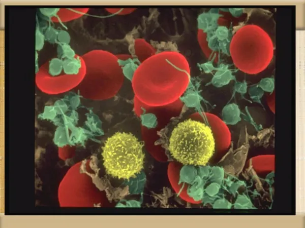
Disorders of Platelets Thrombocytopenia – decreased numbers of platelets (below 100,000/mm3) Can lead to spontaneous bleeding, if low enough, and can be fatal if bleeding occurs in the G.I. Tract, respiratory system or central nervous system. Can be congenital or acquired; acquired is more common. Seen with: Generalized bone marrow suppression Acute viral infection Nutritional deficiencies of B12, folic acid and iron Bone marrow transplant drugs, especially heparin, and toxins, thiazide diuretics, gold, ethanol… Immune reactions 25
Sites of Bleeding in Thrombocytopenia • Skin and mucous membranes • Petechiae • Ecchymosis • Hemorrhagic vesicles • Gingival bleeding and epistaxis • Menorrhagia • Gastrointestinal bleeding • Intracranial bleeding
Quantitative disorders Abnormal distribution Dilution effect Decreased production Increased destruction Qualitative disorders Inherited disorders (rare) Acquired disorders Medications Chronic renal failure Cardiopulmonary bypass Classification of Platelet Disorders
Associated with bleeding Immune-mediated thrombocytopenia (ITP) Most drug-induced thrombocytopenias Most others Associated with thrombosis Thrombotic thrombocytopenic purpura DIC Heparin-associated thrombocytopenia Acquired Thrombocytopenia with Shortened Platelet Survival
Sequestration Hypersplenism Hypothermia Burns Impaired platelet production Aplastic anemia Myelodysplastic syndrome Marrow infiltrative process Osteopetrosis Nutritional deficiency states (iron, folate, viatmin B12, anorexia nervosa) Drug or radiation-induced thrombocytopenia Increased platelet destruction Immune thrombocytopenia Acute and chronic ITP Drug-induced immune thrombocytopenia Post-transfusion purpura Allergy and anaphylaxis Non-immune thrombocytopenia Thrombocytopenia of infection Thrombotic microangiopathic disorders: HUS, TTP Platelets in contact with foreign material Congenital heart disease VWD Combined platelet and fibrinogen consumption syndromes DIC Virus-associated hemophagocytic syndrome Acquired Platelet Defects-Thrombocytopenia
Autoimmune Thrombocytopenia Purpura Idiopathic (primary) By exclusion Dx , no identifiable underlying case Secondary Infection Collagen vascular diseases Lymphoproliferative disorders Solid tumors Drugs Miscellaneous
Features of Acute and Chronic ITP Features Acute ITP Chronic ITP Peak age Children (2-6 yrs) Adults (20-40 yrs) Female:male 1:1 3:1 Antecedent infection Common Rare Onset of symptoms Abrupt Abrupt-indolent Platelet count at presentation <20,000 <50,000 Duration 2-6 weeks Long-term Spontaneous remission Common(80%) Uncommon
Initial Treatment of ITP Platelet count Symptoms Treatment (per µl) >50,000 None 20-50,000 Not bleeding None Bleeding Glucocorticoids IVIG <20,000 Not bleeding Glucocorticoids Bleeding Glucocorticoids IVIG Hospitalization
Practical Aspects for the management of thrombocytopenia What is an adequate platelet count for procedures? Routine Dentistry >10 000 Dental Extraction >30 000 Regional Dental Block >30 000 Minor Surgery >50 000 Major Surgery>80 000 Epidural is okay at platelet count 50 000 for patient with ITP The target platelet count for a bleeding patient is generally >40 000 Prophylactic platelet transfusions for platelets < 10 000
Liver Disease and Hemostasis • Decreased synthesis of II, VII, IX, X, XI, and fibrinogen • Dietary Vitamin K deficiency (Inadequate intake or malabsortion) • Dysfibrinogenemia • Enhanced fibrinolysis (Decreased alpha-2-antiplasmin) • DIC • Thrombocytoepnia due to hypersplenism
Management of Hemostatic Defects in Liver Disease • Treatment for prolonged PT/PTT • Vitamin K 10 mg SQ x 3 days - usually ineffective • Fresh-frozen plasma infusion • 25-30% of plasma volume (1200-1500 ml) • immediate but temporary effect • Treatment for low fibrinogen • Cryoprecipitate (1 unit/10kg body weight) • Treatment for DIC (Elevated D-dimer, low factor VIII, thrombocytopenia • Replacement therapy
Vitamin K deficiency due to warfarin overdoseManaging high INR values Clinical situation Guidelines INR therapeutic-5 Lower or omit next dose; Resume therapy when INR is therapeutic INR 5-9; no bleeding Lower or omit next dose; Resume therapy when INR is therapeutic Omit dose and give vitamin K (1-2.5 mg po) Rapid reversal: vitamin K 2-4 mg po (repeat) INR >9; no bleeding Omit dose; vitamin K 3-5 mg po; repeat as necessary Resume therapy at lower dose when INR therapeutic Chest 2001:119;22-38s (supplement)
Vitamin K deficiency due to warfarin overdoseManaging high INR values in bleeding patients Clinical situation Guidelines INR > 20; serious bleeding Omit warfarin Vitamin K 10 mg slow IV infusion FFP or PCC (depending on urgency) Repeat vitamin K injections every 12 hrs as needed Any life-threatening bleeding Omit warfarin Vitamin K 10 mg slow IV infusion PCC ( or recombinant human factor VIIa) Repeat vitamin K injections every 12 hrs as needed Chest 2001:119;22-38s (supplement)
Hemorrhage in Oral Surgery • Hemorrhage following Oral Surgical procedures can occur due to Local or Systemic causes. • In healthy patients the postoperative bleeding is mainly due to local causes. • Local causes of hemorrhage originate in either Soft Tissue or Bone.
Local causes of hemorrhage in oral surgery –Soft tissue bleeding • Soft tissue bleeding is either arterial, venous, or capillary in nature. • Arterial bleeding is bright red and spurting in nature. • Arteries in the soft tissues at risk during oral surgical procedures are the lies posterior portion of hard palate) greater palatine artery and the buccal artery (lies lateral to the retromolar pad) • Venous blood is dark red in color and flows steadily and heavily especially if the vein is large. • Capillary bleeding is bright red in color and is more of a minimal ooze.
Local causes –Osseous (Bony) bleeding in oral surgery Troublesome bone bleeding originates either from nutrient canals in the alveolar region, central vessels, such as the inferior alveolar artery, or from central vascular lesions (Hemangioma or Vascular malformation)
Systemic causes of hemorrhage in oral surgery • Some patients with heriditary conditions such as hemophilia, Von Willebrand’s disease are susceptible for hemorrhage following oral surgical procedures. • Patients with thrombocytopenia, are at risk of prolonged bleeding after surgery. • Patients with uncontrolled hypertension. • Patients with H/O prosthetic heart valve replacement, Stroke (Cerebrovascular accident) e.t.c., take oral anticoagulants like Aspirin or Warfarin to prevent the occurrence of a thromboembolic episode. • These patients are also at risk of prolonged severe bleeding during and after an oral surgical procedure.
Types of Hemorrhage - Primary Hemorrhage • This occurs during the surgery, as a result of injury like cutting or laceration of the artery or bleeding from bone. • This also occurs when surgery is done in an infected area with a lot of granulation tissue. • It can also occur after a very short period of time immediately after surgery. • This type of bleeding is really normal and can be controlled easily.
Intermediate / Reactionary Hemorrhage • This type of bleeding occurs within a few hours after surgery. • This type of bleeding occurs as a result of failure of coagulation to occur (as in patients with systemic bleeding problems or those on anticoagulants) • Patients who have unknowingly disturbed / dislodged the clot are also prone for this type of bleeding.
Secondary Hemorrhage • This occurs after 7 to 10 days after surgery. This is mainly due to partial division of blood vessel in combination with infection of the wound (Like patient’s who undergo radical neck dissection e.t.c.,). • This type of bleeding is not very frequently encountered after oral surgery procedures.
Management of Primary Hemorrhage in Normal patients The management of bleeding during surgery (Primary bleeding) can be achieved by the following means, • Securing / ligation of blood vessels with silk sutures. • Use of pressure swab to achieve hemostasis. • Use of electrocautery to achieve hemostasis. • Use of hemostatic agents like bone wax, surgicel,e.t.c., • Hypotensive anaesthesia (G.A) and use of vasoconstrictors in L.A.
Local Measures ( Synthetic Materials) • There are several materials that are commercially available that are used locally for achieving adequate hemostasis. • Surgical (Oxidised Regenerated Cellulose) • Gelfoam with activated thrombin • Gelfoam with activated thrombin • Avitene (Microfibrillar Collagen) • Etik Collagen (Packed collagen) • Tranexamic acid 5% • Tranexamic acid 5% in Syringe • Irrigation of wound with Tranexamic acid • Suturing the wound • Pressure with oral packs