Download
1 / 17
1.89k likes | 8.04k Vues
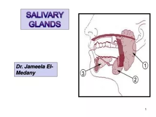
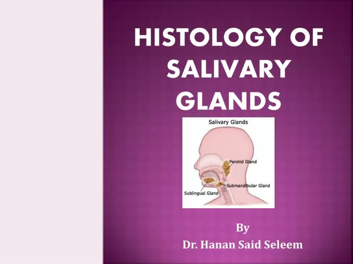
Histology of salivary glands. By Dr. Hanan Said Seleem. Introduction. Three major paired salivary glands: the submandibular , the sublingual and the parotid glands. Exocrine glands that produce saliva. branched tubuloalveolar glands

E N D
Histology of salivary glands By Dr. Hanan Said Seleem
Introduction • Three major paired salivary glands: the submandibular, the sublingual and the parotid glands. • Exocrine glands that produce saliva. • branched tubuloalveolar glands • The minor salivary glands are located in submucosa of different parts of oral cavity.
Structure of salivary glands Salivary gland consists of: • Stroma • parenchyma
The stroma Consists of: • Capsule of CT covering the gland • Trabeculae or septae arise from capsule divide the gland into lobes & lobules • Reticular tissue
The parechyma • Secretory units • Duct system
Secretory units(salivary acini) • Acinus is a group of cells surrounding a lumen • Cells lie on a basement membrane BUT separeted from it by basket cells. • There are three types of acini • Serous acinus • Mucous acinus • Mucoserousacinus
Mixed acinus ( muco-serous): • Mucous acinus which is capped by serous cells forming a serous demilune called crescent of Gianuzzi • Secretion of demilune passes to the lumen through canaliculi between the mucous cells.
Basket cells • Lie between the secretory cells of the acini and the BM. • Contain actin & myosin so called myoepithelial cells • Their contraction squeez the secretion out into the duct system
The duct system Includes: • Intercalated (intercalary) ducts arise from secretory units lined with flat or low cuboidal. • Several intercalated ducts merge with each other to form striated ducts, composed of a single layer of cuboidal to low columnar cells, The basolateral membranes of these cells are highly folded, occupied by mitochondria • Intra- lobular (secretory or salivary) ducts inside the lobules. They take part in secretion. Liend with cubical or columnar cells • Inter-lobular (excretory) ducts in septa between lobules & lined with columnar cells • Inter-lober ducts larger ducts in septa in between lobeslined with psudostratified columnar cells • Main duct union of interlober ducts lined with stratified columnar then st. sq near opening in mouth cavity
Parotid glands • Capsule & septa are thick, fibrous & well developed • Fat cells accumulate around the capsule • Acini are purely serous • Intralobular ducts are very prominent & extensive
Submaxillary (submandibular) • Capsule & septa are thick & fibrous • Fat cells present in septa but less numerous than parotid. • Acini are predominantly serous with some mucous & mixed acini • Intra lobular ducts are very prominent & more extensive than parotid.
Sublingual gland • Capsule is thin & indistinct but septa are well developed • Acini are predominantly mucous with some crescents of Gianuzzi • Intralobular ducts are fewer than thoseof parotid & submaxillary glands.