Download
1 / 29
320 likes | 561 Vues
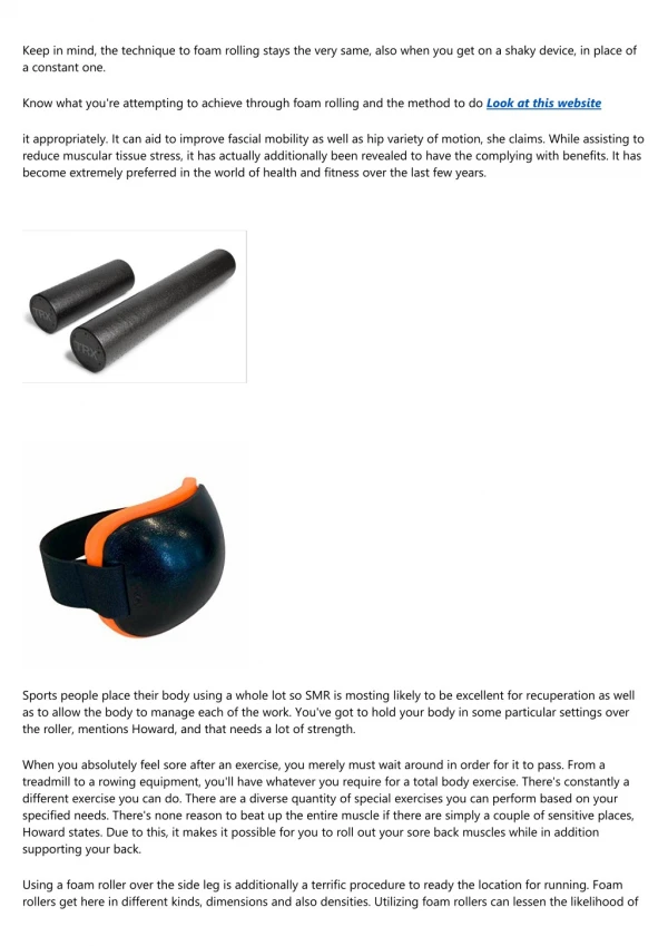
Myofascial Release Techniques for the Lumbopelvic Region. Thomas Cappaert, PhD, ATC, CSCS Central Michigan University GLATA 2010, Detroit, MI . Objectives of Presentation. Review indications for myofascial release. Discuss common dysfunction patterns in the lumbopelvic region.

E N D
Myofascial Release Techniques for the Lumbopelvic Region Thomas Cappaert, PhD, ATC, CSCS Central Michigan University GLATA 2010, Detroit, MI
Objectives of Presentation • Review indications for myofascial release. • Discuss common dysfunction patterns in the lumbopelvic region. • Discuss developing a treatment plan. • Describe common myofascial release techniques. • Demonstrate/practice the techniques.
How does fascia become injured? • Dysfunction comes (secondary to inadequate or over adequate mechanical stress) from the following sources: • ADLs • Work • Leisure/sport • Environment (i.e. furniture, shoes, cars) • Attempts to treat the structure/function continuum • Attempt to identify causative/contributing factors using the “Tight/Loose” concept • Conceptual models of fascia function/dysfunction • Fascia as a balloon • Fascia as plastic wrap • Fascia and muscle as elastic bands
Characteristics of Muscles Relative to “Tight-Loose” Concept
MFR Treatment Indications • Pain complaints have not responded to conservative treatment • Complaints are non-specific to one anatomic structure or region • Underlying chronic condition that leads to soft-tissue tightness • Postural abnormalities • Asymmetrical muscle weakness • ROM has not improved with traditional/conservative treatment
Developing a Treatment Plan • Basic evaluation • Posture • Joint integrity/movement • Signs of dysfunction • Flexibility/strength • Balance/coordination • Local signs/symptoms • Muscle groups involved • Source of dysfunction • Chain reactions • Gross compensations • Developing the plan • What objective information connects to the subjective information? • What’s loose, what’s tight? • What are the asymmetries? • Where are the malalignments?
Common Treatment Sequences • Lateral Hip/Pelvis • Iliac Crest Release • Tensor Fascia Lata Release • Iliotibial Band Release • Quadratus Lumborum Release • Posterior Hip/Pelvis • Sacral Traction • Gluteus Maximus Release • Sacrotuberous Ligament Release • Piriformis Release • Erector Spinae Release • Multifidis Release • Pelvic Roll with Lumbosacral Traction
Common Treatment Sequences continued • Anterior Hip/Pelvis • Quadricep/Anterior Thigh Release • Iliacus Release • Psoas Release • Rectus Abdominus Release • Standing Adductor Release
Practical Applications • Questions? • Demonstration & Practice
Iliac Crest Release • Patient Position • Sidelying with knee flexed and hip flexed to 300. Upper leg is supported by lower leg and spine is in neutral. • Clinician Position • Standing behind patient at waist level and facing toward patient’s feet. • Technique • Use a soft fist or fingers to engage the fascia along the iliac crest. • Start at the midline of the frontal plane and sink the fist inferiorly and produce tension posteriorly and move towards the PSIS as you encounter new layers of tension. • Encourage patient to produce anterior and posterior tilts of the pelvis to uncover additional tension.
Tensor Fascia Lata Release • Patient Position • Sidelying with knee flexed and hip flexed to 300. Upper leg is supported by lower leg and spine is in neutral. • Clinician Position • Standing behind patient at waist level and facing toward patient’s feet. • Technique • Use a soft fist or elbow to engage the muscle just anterior to the gluteus medius. • Engage the initial layers of tension and as tension releases, sink the fist deeper. • When a noticeable change to tone has occurred, add a line of tension inferiorly towards the feet. • Encourage patient to produce anterior and posterior tilts of the pelvis to uncover additional tension.
Iliotibial Band Release • Patient Position • Sidelying with knee flexed and hip flexed to 300. Upper leg is supported by lower leg and spine is in neutral. • Clinician Position • Standing behind patient at waist level and facing toward patient’s feet. Move toward the foot of the table as the release progresses. • Technique • Use a soft fist or elbow to engage the fascia at the greater trochanter. • Engage the initial layers of tension lightly. • When a noticeable change to tone has occurred, add a line of tension inferiorly towards the feet. • Divide the band into sections and repeat the release for each section all the way to tibia. • Work within tolerance levels (if they are visibly in pain or guarding, the work is too deep)
Quadratus Lumborum Release • Patient Position • Sidelying with knee flexed and hip flexed to 300. Upper leg is supported by lower leg and spine is in neutral. • Clinician Position • Standing behind patient at waist level and facing toward patient’s feet. • Technique • Use a soft fist or fingers to engage the muscle just superior to the iliac crest. • Start at the midline of the frontal plane and sink the fist inferiorly towards the transverse processes of the lumbar spine. Increase pressure as you encounter new layers of tension. • Encourage patient to produce anterior and posterior tilts of the pelvis to uncover additional tension. • You may add a posterior line of pressure to also engage the posterior layers of the thoracolumbar fascia.
Sacral Traction • Patient Position • Prone • Clinician Position • Standing at head of patient and facing patients feet. • Technique • Place one hand flat on skin at thoracolumbar junction to stabilize • Place other hand flat on sacrum with heel of hand at lumbosacral junction • Using hand at sacrum, apply tension on an inferior line towards the feet while the hand placed superiorly acts as a counter-force • Continue the inferior tension as tissues release
Gluteus Maximus Release • Patient Position • Prone • Clinician Position • Standing beside the patient at waist level, working on the contralateral side • Technique • Place pads of fingers on both hands at tissue over the PSIS and intermediate iliac crest. • Create a line of tension toward the greater trochanter. • Maintain a consistent depth of pressure as you work laterally. Increase tension as superficial tension dissipates. • Slight anterior and posterior tilts of pelvis will deepen the release.
Sacrotuberous Ligament Release • Patient Position • Prone • Clinician Position • Standing beside the patient at waist level, working on the ipsilateral side • Technique • Using an elbow, fingers or thumb, sink anteriorly through the gluteus maximus. The ligament can be located midway along its attachment to sacrum and 2 cm lateral and inferior to the coccyx. • Create downward/anterior pressure until the ligament is contacted. • Create a line of tension inferiorly toward the ischial tuberosity. • Maintain consistent pressure until tension dissipates and ligament softens. • Slight internal rotation of the ipsilateral leg will deepen the release.
Piriformis Release • Patient Position • place patient side lying with affected leg uppermost and both legs flexed at hip and knee • Clinician Position • Face patient at hip level • Technique • place elbow tip at piriformis insertion (behind greater trochanter) and stabilize pelvis against your trunk • With other hand grasp ankle of affected leg and place into internal rotation to remove slack in the piriformis • Apply moderate pressure with elbow while piriformis is stretched for 5-7 seconds • Then perform an isometric contraction of piriformis (25% of max) for 5-7 seconds • After contraction ceases, take muscle to new barrier and reapply compression with the elbow
Erector Spinae Release • Patient Position • Patient prone with pelvis and feet supported • Clinician Position • Standing to the side at waist level • Technique • use light to moderate, diffuse pressure (soft fist or heel of hand or thumb) at the laminar groove at level of T12 • Treating unilaterally, once tissue slack is removed, add a line of tension inferiorly. • Treat tissue in sections and repeat the release for each section all the way to the sacrum.
Multifidis Release • Patient Position • Patient prone with pelvis and feet supported • Clinician Position • Standing to the side at waist level • Technique • use moderate direct pressure with thumb or finger just lateral to the lumbar spinous processes • Treating unilaterally, once tissue slack is removed, add a line of tension anteriorly. • Treat tissue in sections and repeat the release for each section all the way to the sacrum.
Pelvic Roll with Lumbosacral Traction • Patient Position • Supine with knees flexed and the feet flat on the table. • Clinician Position • Standing beside the patient at mid-thigh level facing toward the head of the table. • Technique • Position at patient with one arm between patients legs resting on the elbow with the forearm supinated and hand resting on the table. • Patient initiates a posterior pelvic tilt and clinician slides hand superiorly so that the hand reaches up under the sacrum. Continue to encourage the pelvic roll, so the hand can be positioned with the fingers at the L1-L2 region with two fingers on each side of the spinous process. • Patient is then instructed to let spine and pelvis rest back fully onto clinicians hand. • Clinician leans on elbow creating a flexion of the fingers and engagement with the tissue. • Clinician then “lifts” through the fingertips and pulls inferiorly towards the feet. • Treat the lumbar spine in sections and carry through treatment to the coccyx.
Quadricep/Anterior Thigh Release • Patient Position • Supine • Clinician Position • Standing at the patients side at hip level • Technique • Use an elbow or soft fist to engage the tissue inferior to the ASIS. Create tension in an inferior direction. Work incrementally toward the knee, dividing the muscle into 3-4 segments. • Abduct the leg to 150. Use fingertips or elbow to sink slowly into the tissue of the femoral triangle in a posterior direction and then create a line of tension in the same direction as the sartorius. • Locate the greater trochanter and the ITB. Using fingertips, palpate for the seam between the ITB and vastus lateralis. Create a line of tension in an inferior direction and treat incrementally. • Have patient perform hip hiking against the line of treatment.
Iliacus Release • Patient Position • Supine with knees supported on bolsters. • Clinician Position • Standing at the patients side at hip level • Technique • Treating one side at a time, use fingertips to locate the medial aspect of the ilium at the ASIS. • Keep the fingerpads touching the bone (with a slight lateral direction) while the tips sink in an inferior/posterior direction. • Engage the first layer of restriction and wait. Once the release occurs, sink to the next layer and continue to treat as appropriate. • Slight posterior pelvic tilt (abdomen drops posteriorly) will improve release.
Psoas Release • Patient Position • Supine with knees supported on bolsters. • Clinician Position • Standing at the patients side at hip level • Technique • Treating one side at a time, locate the psoas by drawing an imaginary line between the umbilicus and the ASIS. • Use the fingers to make contact on this line about halfway between the ASIS and the edge of the rectus abdominus. • Sink in a medial/posterior line and angle in from the lateral edge of the psoas. • Engage the first layer of restriction and wait. Once the release occurs, sink to the next layer and continue to treat as appropriate. • It may take several minutes and a few treatment sessions to get full benefit from the treatment.
Rectus Abdominus Release • Patient Position • Supine with knees extended. • Clinician Position • Standing at the patients side at mid-thigh level • Technique • Treat both sides simultaneously. • Use the fingertips to engage the lateral margins of the rectus abdominus about 2 cm above the pubic bones. Make a “scooping” motion that begins by first sinking posteriorly into the abdominal wall to engage the aponeuroses of the external and internal oblique. • Once the connection is established, lift under the margins of the rectus to put a line of tension in a medial, anterior and superior direction. This completes the scooping action. Maintain this triplanar line of tension and move superiorly. Initially a local stretch will be felt that will progress deeper as tissue relaxes. • Then, span each iliac crest with the fingers and rest the thumbs on the pubic tubercles. Palpate the pubic symphysis. Maintain this contact at the pubic bones and spread the contact through the whole of both hands (not just the thumbs) in a posterior direction. • As the release occurs, bring feet to flat on the table and ask for a posterior pelvic tilt. As the abdomen relaxes and “opens up” release pressure to maintain a lighter contact as the tissue relaxes further.
Standing Adductor Release • Patient Position • Standing with feet about shoulder width apart. • Clinician Position • Kneeling or seated on a stool beside the patient. • Technique • Use the fingerpads of both hands to grasp the adductors about a hand’s width below the ramus of the ishium. Sink into the tissue by pulling toward yourself (laterally) and then creating a line of tension inferiorly. • Have the patient flex the knee to 300 . As they return to standing, maintain the tension in the same line and work as a counterforce to their movement. • Repeat at 2-3 more sites down to the knee.
Resources • Chaitow, L. (2006). Muscle Energy Techniques. (3rd ed.) Edinburgh: Churchill-Livingstone. • Chaitow, L. & S Fritz. (2006). A Massage Therapist’s Guide to Understanding, Locating and Treating Myofascial Trigger Points. Edinburgh: Churchill-Livingstone. • Chaitow, L. (2002). Positional Release Techniques. (2nd ed.) Edinburgh: Churchill-Livingstone. • Manheim, C. (2001). The Myofascial Release Manual. (3rd ed.) Thorofare, NJ: Slack, Inc. • McGill, S. (2006). Ultimate Back Fitness and Performance. (3rd ed.) Waterloo, ON: Backfitpro, Inc. • McGill, S. (2007). Low Back Disorders. (2nd ed.) Champaign, IL: Human Kinetics. • Sahrmann, SA. (2002). Diagnosis and Treatment of Movement Impairment Syndromes. St. Louis, MO: Mosby. • Schamberger, W. (2002). The Malalignment Syndrome. Edinburgh: Churchill-Livingstone. • Schwind, P. (2003). Fascial and Membrane Technique. Edinburgh: Churchill-Livingstone. • Smith, J. (2005). Structural Bodywork. Edinburgh: Churchill-Livingstone. • Stanborough, M. (2004). Direct Release Myofascial Technique. Edinburgh: Churchill-Livingstone.