Bacterial Morphology
Bacterial Morphology. Dr. Abdulaziz Al- Khattaf. Protoplasm. Bacteria organized in units known as cells . Cells are composed of a body protoplast , enclosed by a thin semi-permeable membrane , cytoplasm and a cell-wall .

Bacterial Morphology
E N D
Presentation Transcript
Bacterial Morphology Dr. Abdulaziz Al-Khattaf
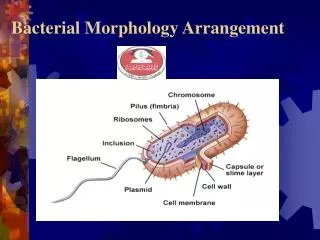
Protoplasm • Bacteria organized in units known as cells. Cells are composed of a body protoplast, enclosed by a thin semi-permeable membrane, cytoplasm and a cell-wall. • Bacteria are microscopic living forms, simple (Prokaryotes) and unicellular in structure. • Bacteria can grow in nutritive solid media to form colonies that are visible to the naked eye.
Prokaryotic cells (Bacteria, Rickettsiae, Chlamydiae and Mycoplasma) distinguishing features are: • Nucleus is homogeneous body with no nuclear membrane- separating it from cytoplasm. • Lacks internal membranes isolating the respiratory and photosynthetic enzymes systems in the specific organelles. • Contains a rigid peptidoglycan cell-wall for support and protection.
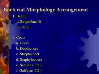
Bacterial shapes • Due to cell-wall presence bacteria could be in spherical (coccus), rod-shaped (bacillus), comma-shaped (vibrio), spiral (spirochaete) or filamentous.
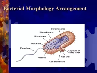
Bacterial Structure • Protoplast: the whole body of living material. • Cytoplasmic membrane: holding and containing the protoplast. • Cell wall: outside rigid supporting the cell (porous-relatively permeable). • Cytoplasm: consist of watery sap containing ribosomes, mesosomes and chromatin. • Inclusion granules: for storage products such aspolyphosphate, lipids and starch.
Capsule: protective gelatinous covering layer. • Flagella: filamentous organs of locomotion. • Fimberiae: organs of adhesion. • Pili: involve in the transfer of genetic material.
Bacterial DNA: • Genetic information of bacterial cell contained in a single, circular double-stranded DNA. • DNA undergoes semi-conservative replication. • Cytoplasm of bacteria: • A viscous watery solution, containing organic and inorganic solutes and ribosomes.
Ribosomes: • They are distributed throughout the cytoplasm and are the sites of protein synthesis. • On the ribosomes subunits (30S and 50S), the mRNA would form peptide sequences. • Transfere RNA (tRNA) molecules would built the peptide sequences into specific polypeptides.
Cytoplasmic inclusions: • Volutin granules (metachromatic granules) Source of stored energy (polymetaphosphate), found in diphtheria bacillus with methylene blue dye or with Albert and Neisser staining. • Lipid-granules: act as carbon and energy storage product. • Polysaccharide granules: either starch or glycogen.
Spores • Spores produced by bacteria in the genera Bacillus and Clostrridium enable them to survive hard environment conditions. • Spores are developed within of vegetative cells. • Spores are resistant to heat, desiccation and disinfectants. • Spores are described as terminal, sub-terminal or central.
Cell Wall • Cell-wall provides rigidity and protects bacterial cell against osmotic damage. • Porous and permeable to substances of low molecule weight. • Structure of cell-wall differs in Gram-positive and Gram-negative. • Gram-negative cell contains an outer membrane with specific proteins that form porins.
Cell Wall • Through these porins hydrophilic molecules are transported. • Other proteins are receptor sites for phages and bacteriocins. • Lipopolysaccharide O antigens and lipid (endotoxin) are embedded in the outer membrane. • Gram-positive cell wall has much thicker layer of peptidoglycan than gram-negative cell-wall. • Teichoic acids are part of the cell wall of gram positive bacteria. They maintain divalent cation outside the cytoplasmic membrane.
Bacteria can survive with defective cell-walls and this was demonstrated in the laboratory with the presence of antibiotics and hyperosmatic environments. • Mycoplasma: independent bacterial genus lacks cell-wall. • L-forms: bacteria with wall-deficient result of penicillin treatment. Can survive and replicate on ordinary media.
External structures • Flagella: Helical filaments, which produce motility by rotation. • Monotrichous is a single polar flagellum; lophotrichous has two or more polar flagella at one end of the cell; amphitrichous has a single flagellum at each end of the cell; and peritrichous with flagella distributed over the cell. • A flagellar protein (H antigen) is useful for helping distinguish between serotvars (serotypes) or variation within a species.
External structures • fimbriae and Pili: many bacteria cells have numerous hairlike structure (fimbriae) that are shorted than flagella. • Fimbriae help the cell to adhere to surfaces such as mucous membranes. • They are often a factor in pathogenicity. • Pili: are les in number than fimbriae (one or two) and called sex pili –they function in transfere of DNA from one cell to another.
External structures • capsules: amorphous (formless) material which surrounds many bacterial species as their outermost layer. • Usually polysaccharide, occasionally protein. • Usually inhibit phagocytosis and their presence correlates with virulence in certain bacteria.
Bacterial Staining • Staining simply coloring the microorganism with dye that emphasizes certain structures. • Fixing (attaching) bacteria to the slide is the first step. • Stains are salt compose of a positive or negative ions. Basic dyes, are positively ion charged where, in acidic dyes, negatively ion charged. • Bacteria cell is slightly negatively charged so attracting basic dyes (crystal violet, methylene blue, malachite green, and safranin).
Bacterial staining • Acidic dyes: are not attracted to bacteria because of its negative charge that repelled by the bacterial negative charge. • This stain colors the background instead (negative staining). • This is useful to observe the over all cell shape, sizes and capsules (eosin, acid fuchsin and nigrosin).