Download
1 / 44
470 likes | 837 Vues
Upper Respiratory System Digital Laboratory. It’s best to view this in Slide Show mode, especially for the quizzes. This module will take approximately 75 minutes to complete . After completing this exercise, you should be able to:

E N D
Upper Respiratory System Digital Laboratory It’s best to view this in Slide Show mode, especially for the quizzes. This module will take approximately 75 minutes to complete.
After completing this exercise, you should be able to: • Describe the general features of structures in the upper respiratory tract: • Respiratory epithelium • Basement membrane (thick) • Lamina propria with glands, vessels, nerves, diffuse lymphoid tissue • Supporting elements (bone, cartilage, smooth and/or skeletal muscle • Distinguish, at the light microscope level, each of the following organs and their specific features (in addition to those listed above): • Concha – venous plexus , cancellous bone • Epiglottis - epithelium (stratified squamous non keratinized on anterior side and tip, respiratory epithelium on posterior side), elastic cartilage • Larynx – • False vocal fold – mucus and serous glands • True vocal fold - stratified squamous epithelium, vocal ligament , vocalis muscle, lack of blood vessels • Laryngeal ventricle • Hyoid bone • Epiglottis • Thyroid cartilage • Cricoid cartilage • Trachea – submucosa with glands, C-shaped hyaline cartilage , trachealis muscle • Review, at the electron microscopic level, respiratory epithelium and cilia
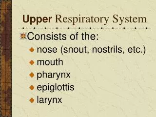
GROSS ANATOMY OF THE UPPER RESPIRATORY TRACT • Look and learn. Note: • Nasal cavity • Pharynx (studied with digestive tract) • Larynx • Trachea • Bronchi Different sources define upper vs. lower respiratory systems differently. Clinicians use these terms to describe upper respiratory infections (colds) and lower respiratory infections (pneumonia). Based on histological similarity, we will address everything up to and including the bronchi as upper respiratory., and will focus on structures in the lungs in the next laboratory.
RESPIRATORY MUCOSA • The inner lining of the respiratory system is largely composed of respiratory mucosa, which includes • Pseudostratified ciliated columnar epitheium with goblet cells • Lamina propria– irregular connective tissue with glands and diffuse lymphoid tissue The supporting structures of the respiratory system (bone, cartilage, smooth muscle) will be addressed when we examine specific regions of the respiratory system.
RESPIRATORY MUCOSA • The inner lining of the respiratory system is largely composed of respiratory mucosa, which includes • Pseudostratified ciliated columnar epitheium with goblet cells • Thick basement membrane (green bracket) • Lamina propria– irregular connective tissue with glands and diffuse lymphoid tissue
RESPIRATORY MUCOSA • The inner lining of the respiratory system is largely composed of respiratory mucosa, which includes • Pseudostratified ciliated columnar epitheium with goblet cells • Thick basement membrane • Lamina propria– irregular connective tissue with glands and diffuse lymphoid tissue • Remind yourself of the ultrastructural features of cilia: • 9+2 arrangement of microtubules • Basal bodies.
QUIZ Self-check: Identify 1-6. (advance slide for answers)
QUIZ Self-check: Identify 1-6. (advance slide for answers)
QUIZ Self-check: Identify 1-7. (advance slide for answers)
CONCHAE • The upper respiratory tract is designed to filter, warm, and moisturize incoming air. • The nasal cavity is well suited for this function because it contains: • Respiratory epithelium • Numerous venous plexuses • Projections of the lateral wall of the nasal cavity are called conchae. These structures increase turbulence within the nasal cavity, enhancing it’s function of treating air. The histological features of the conchae are similar to most of nasal cavity.
CONCHA The next video shows a concha similar to the one outlined in green.
CONCHA Video showing concha – slide 110 • Link to SL 110 • Be able to identify: • Concha • Mucosa • Respiratory epithelium • Thick basement membrane • Lamina propria • Glands • Venous plexus • Bone
EPIGLOTTIS The epiglottis helps guide swallowed food toward the esophagus by covering the opening into the larynx. The drawing below shows the epiglottis in the closed position (left) and open position (right). The supporting element of the epiglottis is elastic cartilage, which is firm but flexible.
EPIGLOTTIS The anterior side and the posterior tip of the epiglottis (surfaces colored in green) contact food and require a stratified squamous epithelium to resist friction. The remainder of the posterior surface (colored orange) does not encounter friction, and is covered by respiratory epithelium • Next we’ll look at two digital slides of the epiglottis: • The first slide of the epiglottis is a cross section (yellow line) of the tip, stained for elastic fibers. • The second slide is a sagittal cut, similar to the plane of this drawing (with the tip toward the left).
EPIGLOTTIS Video showing cross section of epiglottis, elastic stain – slide 184 • Link to SL 184 • Be able to identify: • Epiglottis • Elastic cartilage
EPIGLOTTIS Video showing longitudinal section of epiglottis – slide 17 • Link to SL 017 • Be able to identify: • Epiglottis • Epithelium • Stratified squamous • Respiratory • Lamina propria • Connective tissue, nerves, vessels, serous and mucus glands • Elastic cartilage
LARYNX The larynx is positioned anterior to the esophagus, and is the gateway for inspired air to enter the trachea and the rest of the respiratory tract.
LARYNX The larynx is supported by the hyoid bone, cartilage, and supporting ligaments. Study these views of the supporting elements: Hyoid bone epiglottis thyroid cartilage cricoid cartilage vestibular ligament vocal ligament
LARYNX The next several drawings show are gross depictions of the larynx. In this drawing of the posterior aspect of the pharynx, the posterior wall of the pharynx and esophagus has been opened, showing you the larynx (yellow box). Clemente 893
LARYNX In this drawing, the posterior wall of the larynx has been opened. Note: vocal fold vestibular fold epiglottis hyoid bone muscles of the larynx Clemente 907 Terminology alert: the vocal and vestibular ligaments are ligaments, when they are covered with mucosa, the entire structure (ligament + mucosa) is referred to as a fold (e.g. vocal fold).
LARYNX B This is a sagittal section through the larynx, anterior is to the left, the esophagus (not shown) is to the right. Note: epiglottis vestibular fold vocal fold laryngeal ventricle thyroid cartilage cricoid cartilage The next drawing and the digital slide are coronal sections, similar to that represented by the dotted yellow line. Clemente 909
LARYNX Coronal section similar to the digital slide on the next screen. Note: epiglottis hyoid bone thyroid cartilage cricoid cartilage muscles vestibular ligament (within fold) laryngeal ventricle vocal ligament (within fold) Clemente 908
LARYNX Video overviewing larynx – slide 36B • Link to SL 036B • Be able to identify: • Larynx • Epiglottis • Thyroid cartilage • Cricoid cartilage • Vestibular fold • Vocal fold • Laryngeal ventricle
LARYNX Video detailing larynx – slide 36B • Link to SL 036B • Be able to identify: • Larynx • Epiglottis – infant elastic cartilage (do not have to ID infant cartilage for exam • Thyroid cartilage –hyaline cartilage • Cricoid cartilage – hyaline cartilage • Skeletal muscle • Peripheral nerve
LARYNX Video detailing vestibular and vocal folds – slide 36B • Link to SL 036B • Be able to identify: • Larynx • Vestibular fold • Respiratory epithelium • Glands • Vocal fold • Stratified squamous epithelium • No glands • Vocal ligament • Vocalis muscle • Laryngeal ventricle
LARYNX The next slide is an oblique cut through the larynx, and shows only one side:
LARYNX Video showing oblique cut through larynx – slide 36A • Link to SL 036A • Be able to identify: • Larynx • Vestibular fold • Respiratory epithelium • Glands • Vocal fold • Stratified squamous epithelium • No glands • Vocal ligament • Vocalis muscle • Laryngeal ventricle
TRACHEA The trachea is a single tube passing inferiorly from the larynx until it bifurcates into the primary bronchi. The trachea is supported by about a dozen C-shaped pieces of cartilage, which are open posteriorly. The trachialis muscle (smooth muscle) spans the open, posterior end of the cartilages.
TRACHEA Video of trachea – slide 15B • Link to SL 015B • Be able to identify: • Trachea • Mucosa • Respiratory epithelium • Lamina propria • Submucosa • Glands • C-shaped hyaline cartilage • Trachealis muscle • Adventitia • Review elastic artery, peripheral nerve, lymph node, unilocular adipose, skeletal and smooth muscle, stratified squamous non-keratinized epithelium
The next set of slides is a quiz for this module. Before continuing, we suggest you remind yourself of the list of objectives covered in this module, and mentally visualize what each region or structure will look like: • Describe the general features of structures in the upper respiratory tract: • Respiratory epithelium • Basement membrane (thick) • Lamina propria with glands, vessels, nerves, diffuse lymphoid tissue • Supporting elements (bone, cartilage, smooth and/or skeletal muscle • Distinguish, at the light microscope level, each of the following organs and their specific features (in addition to those listed above): • Concha – venous plexus , cancellous bone • Epiglottis - epithelium (stratified squamous non keratinized on anterior side and tip, respiratory epithelium on posterior side), elastic cartilage • Larynx – • False vocal fold – mucus and serous glands • True vocal fold - stratified squamous epithelium, vocal ligament , vocalis muscle, lack of blood vessels • Laryngeal ventricle • Hyoid bone • Epiglottis • Thyroid cartilage • Cricoid cartilage • Trachea – submucosa with glands, C-shaped hyaline cartilage , trachealis muscle • Review, at the electron microscopic level, respiratory epithelium and cilia
QUIZ Self-check: Identify 1-6. (advance slide for answers)
QUIZ Self-check: Identify 1-5. (advance slide for answers)
QUIZ Self-check: Identify 1-6. (advance slide for answers)
QUIZ Self-check: Identify the structure / organ from which these images were taken. (advance slide for answers) epiglottis
QUIZ Self-check: Identify the structure / organ from which these images were taken. (advance slide for answers) concha
QUIZ Self-check: Identify the outlined structures. (advance slide for answers) Vestibular fold Vocal ligament
QUIZ Self-check: Identify the structure / organ from which these images were taken. (advance slide for answers) Urinary bladder
QUIZ Self-check: Identify the structure / organ from which these images were taken. (advance slide for answers) trachea