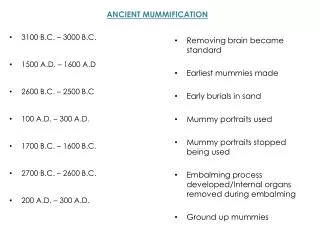

The Ebers papyrus 2600 B.C.
E N D
Presentation Transcript
3. 22:21 3 8 million patients with chest pain present annually to emergency departments. Five million of this group are judged to have suspected acute coronary syndromes and are admitted to the hospital. Less than half ultimately are found to have a cardiac diagnosis.
8 million patients with chest pain present annually to emergency departments. Five million of this group are judged to have suspected acute coronary syndromes and are admitted to the hospital. Less than half ultimately are found to have a cardiac diagnosis.
4. This slide reviews the anatomy of the AV node.
The AV node is divided into two regions, the compact AV node and AV nodal tracts. The compact AV node is a button-like portion of the AV node from which the His bundle originates. AV nodal tracts are bands of conductive tissue that gather electrical impulses from the internodal pathways and deliver them to the compact AV node. The AV nodal tracts that converge to form the compact AV node are poorly defined�that is, it is not entirely clear where the internodal pathways end and the AV nodal tracts begin. Thus far, two distinct tracts have been identified:
Anterior tract (also called the fast AV nodal pathway)
Posterior tract (also called the slow AV nodal pathway)
This picture also illustrates the triangle of Koch, and anatomical landmark used to visualize the location of the AV nodal tracts. The three sides of the Koch�s triangle are defined by the tricuspid valve annulus, the tendon of Todaro, and the ostium of the coronary sinus. The fast AV nodal pathway lies near the compact AV node along the tendon of Todaro. The slow AV nodal pathway is located along the tricuspid valve annulus near the ostium of the coronary sinus.
This slide reviews the anatomy of the AV node.
The AV node is divided into two regions, the compact AV node and AV nodal tracts. The compact AV node is a button-like portion of the AV node from which the His bundle originates. AV nodal tracts are bands of conductive tissue that gather electrical impulses from the internodal pathways and deliver them to the compact AV node. The AV nodal tracts that converge to form the compact AV node are poorly defined�that is, it is not entirely clear where the internodal pathways end and the AV nodal tracts begin. Thus far, two distinct tracts have been identified:
Anterior tract (also called the fast AV nodal pathway)
Posterior tract (also called the slow AV nodal pathway)
This picture also illustrates the triangle of Koch, and anatomical landmark used to visualize the location of the AV nodal tracts. The three sides of the Koch�s triangle are defined by the tricuspid valve annulus, the tendon of Todaro, and the ostium of the coronary sinus. The fast AV nodal pathway lies near the compact AV node along the tendon of Todaro. The slow AV nodal pathway is located along the tricuspid valve annulus near the ostium of the coronary sinus.
6. 22:21 6
7. GaDget para el DOLOR TOR�CICO
8. HERRAMIENTA para manejar el DT
9. GADGET 1: signos de gravedad inminente
GADGET 2 : 5 causas potenciales de muerte
GADGET 3 : probabilidad de enfermedad coronaria
GADGET 4 : habilidad con el ECG
GADGET 5 : estratificaci�n de riesgo
14. HERRAMIENTA para manejar el DT
15. GaDget
16. 22:21 16
18. �Cu�les son sus causas? 1 - Causas m�sculo-esquel�ticas o de origen en la pared tor�cica:
Costocondritis, Roturas o desgarros musculares.
2 - Causas cardiol�gicas:
angina de pecho o SCA
Pericarditis: inflamaci�n del pericardio.
3 - Causas vasculares:
Embolismo pulmonar, Disecci�n de aorta
4 - Causas pulmonares y pleurales:
Neumon�as, Pleuritis, Tumores pulmonares, Neumot�rax
5 - Causas digestivas:
Esofagitis / Hernia de hiato, Espasmos esof�gicos, Rotura esof�gica
6 - Causas mediast�nicas:
La inflamaci�n de esta zona o la lesi�n de las estructuras que por ella discurren puede originar dolor.
7 - Causas neurol�gicas:
Herpes zoster intercostal
8 - Causas psic�genas:
Dolor subjetivo sin que exista lesi�n significativa de ninguno de los �rganos internos y que se produce asociado a cuadros de ansiedad.
22. CLASIFICACIONES DE DOLOR TOR�CICO DEL ESTUDIO CASS Y DE DIAMOND Y FORRESTER
23. 22:21 23
24. GaDget
28. This slide reviews the anatomy of the AV node.
The AV node is divided into two regions, the compact AV node and AV nodal tracts. The compact AV node is a button-like portion of the AV node from which the His bundle originates. AV nodal tracts are bands of conductive tissue that gather electrical impulses from the internodal pathways and deliver them to the compact AV node. The AV nodal tracts that converge to form the compact AV node are poorly defined�that is, it is not entirely clear where the internodal pathways end and the AV nodal tracts begin. Thus far, two distinct tracts have been identified:
Anterior tract (also called the fast AV nodal pathway)
Posterior tract (also called the slow AV nodal pathway)
This picture also illustrates the triangle of Koch, and anatomical landmark used to visualize the location of the AV nodal tracts. The three sides of the Koch�s triangle are defined by the tricuspid valve annulus, the tendon of Todaro, and the ostium of the coronary sinus. The fast AV nodal pathway lies near the compact AV node along the tendon of Todaro. The slow AV nodal pathway is located along the tricuspid valve annulus near the ostium of the coronary sinus.
This slide reviews the anatomy of the AV node.
The AV node is divided into two regions, the compact AV node and AV nodal tracts. The compact AV node is a button-like portion of the AV node from which the His bundle originates. AV nodal tracts are bands of conductive tissue that gather electrical impulses from the internodal pathways and deliver them to the compact AV node. The AV nodal tracts that converge to form the compact AV node are poorly defined�that is, it is not entirely clear where the internodal pathways end and the AV nodal tracts begin. Thus far, two distinct tracts have been identified:
Anterior tract (also called the fast AV nodal pathway)
Posterior tract (also called the slow AV nodal pathway)
This picture also illustrates the triangle of Koch, and anatomical landmark used to visualize the location of the AV nodal tracts. The three sides of the Koch�s triangle are defined by the tricuspid valve annulus, the tendon of Todaro, and the ostium of the coronary sinus. The fast AV nodal pathway lies near the compact AV node along the tendon of Todaro. The slow AV nodal pathway is located along the tricuspid valve annulus near the ostium of the coronary sinus.
29. 22:21 29
30. 22:21 30
31. This slide reviews the anatomy of the AV node.
The AV node is divided into two regions, the compact AV node and AV nodal tracts. The compact AV node is a button-like portion of the AV node from which the His bundle originates. AV nodal tracts are bands of conductive tissue that gather electrical impulses from the internodal pathways and deliver them to the compact AV node. The AV nodal tracts that converge to form the compact AV node are poorly defined�that is, it is not entirely clear where the internodal pathways end and the AV nodal tracts begin. Thus far, two distinct tracts have been identified:
Anterior tract (also called the fast AV nodal pathway)
Posterior tract (also called the slow AV nodal pathway)
This picture also illustrates the triangle of Koch, and anatomical landmark used to visualize the location of the AV nodal tracts. The three sides of the Koch�s triangle are defined by the tricuspid valve annulus, the tendon of Todaro, and the ostium of the coronary sinus. The fast AV nodal pathway lies near the compact AV node along the tendon of Todaro. The slow AV nodal pathway is located along the tricuspid valve annulus near the ostium of the coronary sinus.
This slide reviews the anatomy of the AV node.
The AV node is divided into two regions, the compact AV node and AV nodal tracts. The compact AV node is a button-like portion of the AV node from which the His bundle originates. AV nodal tracts are bands of conductive tissue that gather electrical impulses from the internodal pathways and deliver them to the compact AV node. The AV nodal tracts that converge to form the compact AV node are poorly defined�that is, it is not entirely clear where the internodal pathways end and the AV nodal tracts begin. Thus far, two distinct tracts have been identified:
Anterior tract (also called the fast AV nodal pathway)
Posterior tract (also called the slow AV nodal pathway)
This picture also illustrates the triangle of Koch, and anatomical landmark used to visualize the location of the AV nodal tracts. The three sides of the Koch�s triangle are defined by the tricuspid valve annulus, the tendon of Todaro, and the ostium of the coronary sinus. The fast AV nodal pathway lies near the compact AV node along the tendon of Todaro. The slow AV nodal pathway is located along the tricuspid valve annulus near the ostium of the coronary sinus.
32. 22:21 32
33. Estratificaci�n de riesgo
37. This slide reviews the anatomy of the AV node.
The AV node is divided into two regions, the compact AV node and AV nodal tracts. The compact AV node is a button-like portion of the AV node from which the His bundle originates. AV nodal tracts are bands of conductive tissue that gather electrical impulses from the internodal pathways and deliver them to the compact AV node. The AV nodal tracts that converge to form the compact AV node are poorly defined�that is, it is not entirely clear where the internodal pathways end and the AV nodal tracts begin. Thus far, two distinct tracts have been identified:
Anterior tract (also called the fast AV nodal pathway)
Posterior tract (also called the slow AV nodal pathway)
This picture also illustrates the triangle of Koch, and anatomical landmark used to visualize the location of the AV nodal tracts. The three sides of the Koch�s triangle are defined by the tricuspid valve annulus, the tendon of Todaro, and the ostium of the coronary sinus. The fast AV nodal pathway lies near the compact AV node along the tendon of Todaro. The slow AV nodal pathway is located along the tricuspid valve annulus near the ostium of the coronary sinus.
This slide reviews the anatomy of the AV node.
The AV node is divided into two regions, the compact AV node and AV nodal tracts. The compact AV node is a button-like portion of the AV node from which the His bundle originates. AV nodal tracts are bands of conductive tissue that gather electrical impulses from the internodal pathways and deliver them to the compact AV node. The AV nodal tracts that converge to form the compact AV node are poorly defined�that is, it is not entirely clear where the internodal pathways end and the AV nodal tracts begin. Thus far, two distinct tracts have been identified:
Anterior tract (also called the fast AV nodal pathway)
Posterior tract (also called the slow AV nodal pathway)
This picture also illustrates the triangle of Koch, and anatomical landmark used to visualize the location of the AV nodal tracts. The three sides of the Koch�s triangle are defined by the tricuspid valve annulus, the tendon of Todaro, and the ostium of the coronary sinus. The fast AV nodal pathway lies near the compact AV node along the tendon of Todaro. The slow AV nodal pathway is located along the tricuspid valve annulus near the ostium of the coronary sinus.
39. 22:21 39
40. HERRAMIENTA para manejar el DT
42. This slide shows the rise and fall of CK, CK-MB, cTnI and myoglobin after an AMI.
Note that with the newer second generation cTnI assays, increases are often seen as early as those in myoglobin, and earlier than increases in CK or CK-MB.This slide shows the rise and fall of CK, CK-MB, cTnI and myoglobin after an AMI.
Note that with the newer second generation cTnI assays, increases are often seen as early as those in myoglobin, and earlier than increases in CK or CK-MB.
43. 22/08/2012 43 �� Pron�stico excelente en los pacientes con marcadores negativos, pod�an ser dados de ALTA !!