Download
1 / 1
10 likes | 255 Vues
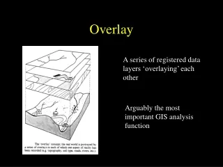
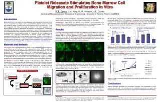
Bone Interface Group. Fibrin Gel Overlay. A: Leading Front of Migration ( m). B: Number of Migrating Cells. p < 0.05. p < 0.05. Control PR 5 PR 5,7 PR 7 Control PR 5 PR 5,7 PR 7 . e. b. c. d. a. f. h. i. j. g.

E N D
Bone Interface Group Fibrin Gel Overlay A: Leading Front of Migration (m) B: Number of Migrating Cells p < 0.05 p < 0.05 Control PR 5 PR 5,7 PR 7 Control PR 5 PR 5,7 PR 7 e b c d a f h i j g Cell Proliferation Total cell number (x1000) 3-D Fibrin Gel Migration Assay Day 10: Cross-section through fibrin gel (F-actin stain) Day 1: Rat bone marrow derived cell subculture 1 3 5 7 10 Day 5: Platelet releasate Days after subculture Cell migration proceeded counter to gravity in response to added growth factors. Cell culture media Fibrin Gel Confluent cell layer Bone nodule formation as observed by light microscopy. Platelet Releasate Stimulates Bone Marrow Cell Migration and Proliferation In Vitro W.E. Oprea, J.M. Karp, M.M. Hosseini, J.E. Davies Institute of Biomaterials and Biomedical Engineering, University of Toronto, Toronto CANADA analyzed by confocal microscopy. Transmission electron microscopy (TEM) was used to verify the presence of de novo bone formation within the fibrin gels. Proliferation: Cells plated at a density of 104 cells/cm2 were subjected to the effects of PR or rhPDGF-BB at parallel time points to the 3-D migration studies. For counting, cells were trypsinized and counted using a hemocytometer. Results Confocal microscopy revealed extensive bone marrow cell infiltration into the fibrin matrix (Figure 1). Determination of the leading front of migration showed migration ranging from 50 to 350m (equivalent to 70 m/day), which corresponds well with published in vivo data [4]. TEM analysis (Figure 2) showed the presence of electron dense bodies containing hydroxyapatite within the fibrin gels, thus confirming that a percentage of the migratory cells were indeed osteogenic. Figure 1.Series of horizontal images obtained by confocal microscopy, depicting cell infiltration into the fibrin gel. Image series begins at bottom of the culture dish (a) where a confluent cell layer is visible, and proceeds up through the unstained fibrin gel. Cells are stained green for F-actin, rending the cell body and extended cell processes clearly visible. Vertical distance between images: 15m. Field width: 265 m. Figure 2.(A) TEM photomicrograph depicting the branch of an osteogenic cell (oC) embedded within the extracellular fibrinous matrix (efM). In proximity to the cell branch, several spherical, electron dense bodies on the order of 0.5 m are visible (arrows), which at higher magnification (B) are shown to contain individual needle-shaped particles (arrows). (C) The presence of hydroxyapatite these foci of mineralization (FM) was confirmed by the circular electron diffraction pattern [5]. Field width A: 10 m. Field width B: 1.7 m. PR was shown to stimulate the migration of RBMC within the 3-D fibrin matrices. In groups with added PR, a 25% increase in the leading front of migration and a 3.5x increase in the number of migrating cells (Figure 3) was observed. Similar trends were observed when rhPDGF-BB was added (not shown); however, the rhPDGF-BB groups were not statistically significant from the control groups. Figure 3.(A) Average leading front of migration, or furthest distance migrated by any one cell at analyzed point in m. (B) Number of migrating cells in the volume above a ~250 x 250 m area. Experimental groups are divided according to day(s) after subculture on which PR was added. Proliferation studies (Figure 4) further demonstrated that PR is mitogenic for RMBC. Comparison between migration and proliferation data indicated that PR also stimulates the recruitment of RBMC to migration. Figure 4: RBMC proliferation. Growth factors were added at parallel time points to the 3-D migration studies. Conclusions Platelet releasate stimulates the recruitment, migration and proliferation of bone marrow derived cells. Our results provide a potential mechanism to explain why biologically active platelet-derived factors may enhance endosseous wound healing. Introduction Before placement of an implant at an endosseous site, extravasated blood will fill the wound compartment and begin to clot. Concomitantly, platelets become activated and release a library of growth factors and cytokines within the injury site. It has been suggested that these factors play an important role in the regulation of the wound healing cascade by stimulating the migration and proliferation of osteogenic cells within the fibrin clot of the injury compartment. Such thinking has encouraged new clinical strategies that incorporate various growth factors with materials in order to accelerate the wound healing process [1]. However, for such strategies to be successful, the underlying mechanisms of platelet growth factor action at endosseous sites must be more completely understood. To date, no information on the effects of platelet factors on bone cell migration is available [2,3]. The purpose of this work was to develop an in vitro model to study the effects of the growth factors contained in platelet releasate (PR) on osteogenic cell migration and proliferation. Materials and Methods Cell culture: Rat bone marrow cells (RBMC) were harvested from the femurs of young Wistar rats and cultured in fully supplemented media (FSM) consisting of -minimal essential media supplemented with 15%(v/v) fetal calf serum, 10% (v/v) antibiotics, 50 g/ml L-ascorbic acid, 5mM -glycerophosphate and 10-8 M dexamethazone. Cultures were incubated at 37°C and 100% relative humidity. Platelet concentrate (PC) was obtained as prescribed by the 3i PCCS ™ system. To obtain PR, PC was activated with 1 IU human thrombin/ml and the supernatant containing the released platelet factors was collected after centrifugation at 2000xg. 3-D Migration: Confluent RMBC cultures in 35 mm tissue culture dishes were overlaid with a 1.5 mm thick fibrin gel, obtained by mixing 3 mg/ml human fibrinogen with 1.5 IU/ml thrombin. These groups were supplemented with 2 ml FSM containing PR (1:4 v/v) or rhPDGF-BB (20 ng/ml) at various time points. On day 5 after gel overlay, cultures were fixed and stained with phalloidin, an F-actin stain, and Acknowledgements The authors gratefully acknowledge the provision of fibrin from Haemacure Corp., and financial support from both Implant Innovations Inc. and an Ontario Research and Development Challenge Fund (ORDCF) grant to JED. References 1. Marx RE et al. (1998) Oral Surg Oral Med Oral Pathol Oral Radiol Endod, 85(6): 683 –646. 2. Haynesworth SE et al. (2002) Poster. ORS, Dallas. 3.Gruber R, et al. (2002) Clin Oral Impl Res. 13: 529-535. 4. Winet et al. (1990) Calcif Tissue Int. 47:24-34. 5. Tennenbaum et al. (1986). Bone. 7: 295-302.