Chest Pain Epidemiology
780 likes | 1.27k Vues
Chest Pain Epidemiology. 6 million ED visits/year 5-7% ED patients 3.3% AIS evacuations 2002, 3.5% in 2003, 3.6% in 2004, 3.2% in 2005 3 million patients admitted/year 70% found not to have acute coronary event 0.4% - 4.0% acute MI are sent home. Chest Pain Pathophysiology.

Chest Pain Epidemiology
E N D
Presentation Transcript
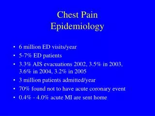
Chest PainEpidemiology • 6 million ED visits/year • 5-7% ED patients • 3.3% AIS evacuations 2002, 3.5% in 2003, 3.6% in 2004, 3.2% in 2005 • 3 million patients admitted/year • 70% found not to have acute coronary event • 0.4% - 4.0% acute MI are sent home
Chest PainPathophysiology • Chest pain syndromes difficult to diagnose • Multiple organ systems of the chest • Share afferent (nerve) pathways • Pathology in any of these systems have similar pattern of complaints • Most patients have CP with acute coronary syndrome(ACS), others may present with only SOB, N/V, arm or jaw pain
Differential Diagnosisof Chest Pain Life-threatening causes • Acute coronary syndrome(ACS) • Aortic dissection • Pulmonary embolism • Tension pneumothorax • Esophageal rupture (Boerhaave’s syndrome) • Pericarditis; myocarditis • Acute chest syndrome(in sickle cell disease)
Differential Diagnosisof Chest Pain Non-life-threatening causes • Gastrointestinal Biliary colic (cholelithiasis, cholecystitis) Gastroesophageal reflux disease Peptic ulcer disease • Pulmonary Pneumonia Pleurisy
Differential Diagnosisof Chest Pain Non-life-threatening causes • Chest wall syndromes Musculoskeletal pain Costochondritis Thoracic radiculopathy • Psychiatric Anxiety • Shingles
Chest Pain Evaluation • Problems • History • Risk factors • Physical exam • Rhythm strip, 9 lead ECG, 12 lead ECG • Risk stratification based on above factors
The Initial Clinical Examination • ECG can only help if it shows acute MI • Initial ECG sensitivity 20% - 60% AMI • Sensitivity of plasma CK-MB low first 4 hrs • Can’t detect unstable angina • Therefore evaluation based on history, physical exam and ECG
History “The most important difference between a good and indifferent clinician lies in the amount of attention paid to the story of the patient”---Farquhar Buzzard
History • Helpful to group questions to target the three most common life threats; Consider ACS questions Pulmonary embolism(PE) questions Aortic dissection questions
HistoryCardiac Questions • 2 most important historical information age, gender • Advancing age, prevalence and severity of CAD increases • Can estimate pretest probability of CAD based on age and gender • Further refine pretest probability by classifying the chest pain as typical, atypical, or non-anginal
Pretest likelihood of CAD based on age, sex, and symptoms Asymptomaticnon-anginal CP Age Men Women Men Women 30-39 1.9% 0.3% 5.2% 0.8% 40-49 5.5% 1.0% 14.1% 2.8% 50-59 9.7% 3.2% 21.5% 8.4% 60-69 12.3% 7.5% 28.1% 18.6%
Pretest likelihood of CAD based on age, sex, and symptoms Atypical angina Typical angina Age Men Women Men Women 30-39 21.8% 4.2% 69.7% 25.8% 40-49 46.1% 13.3% 87.3% 55.2% 50-59 58.9% 32.4% 92.0% 79.4% 60-69 67.1% 54.4% 94.3% 90.6%
Cardiac Questions • Example; 35y/o male with non-anginal CP has 5% pretest probability of CAD(1 in 20) same 35y/o with atypical angina 22% of CAD or (1in 5) same 35y/o with typical angina 70%(7in10) • If patient has known previous CAD/MI raises risk of subsequent coronary event 5 times • If patient has cardiac history ask about prior stress tests, cardiac caths, bypass surgery, stents
Cardiac Questions • Character of Pain • Many patients have atypical symptoms • Ask questions in regard to nature (quality), severity(1-10), duration, modifying factors of the pain, and associated symptoms • 40% patients with AMI have atypical CP • 35% patients without AMI have typical CP
Cardiac Questions • In one study of 721 patients who were diagnosed with AMI, almost ½ presented without CP • SOB, weakness, dizziness, syncope, abdominal pain • Typical angina is a deep, poorly localized chest or arm discomfort that is classically exertional and relieved with rest or nitrates
Analysis of Clinical Predictors of AMI • Clinical features AMI chest pain radiation Odds ratio left arm 1.5 right arm 3.2 both arms 7.7 nausea, vomiting 1.8 diaphoresis 1.4 exertional CP 3.1
Analysis of Clinical Predictors of AMI • Clinical features AMI Odds ratio burning/indigestion pain 4.0 crushing/squeezing pain 2.1 relief with nitroglycerin 0.9 pleuritic pain 0.5 tender chest wall 0.2 sharp/stabbing pain 0.5
Cardiac Questions • Another study of 251 patients with cardiac CP showed 88% respond to NTG, also 92% of noncardiac CP responded to NTG • Can you give GI cocktail to R/O cardiac CP? a study of 97 patients who received GI cocktail showed 8 of 11 patients admitted with possible cardiac ischemia had complete or partial relief of CP
Cardiac Questions • Risk Factors • Diabetes, hypertension, smoking, high cholesterol, and family history • Most CAD patients have at least one • The absence of risk factors does not exclude acute cardiac ischemia
Aortic DissectionHistory • Male (75%) • Seventh decade • History of hypertension (70%) • Other risk factors; Marfan’s syndrome, atherosclerosis, prior dissection, or known aortic aneurysm
Aortic DissectionHistory • Pain is sudden onset (83%) • Severe or “worse ever” (90%) • Sharp (64%) or tearing (50%) • Location anterior chest (60%), back (53%) • Migratory (16%), radiating (28%) • Suspect dissection in patients with clinical changing picture
Aortic Dissection History • Should address 3 basic concerns regarding a patient’s pain: quality (sudden and severe) radiation (especially to the back) intensity at onset (maximal) • Aortic dissection and MI can coexist 8% dissection involves coronary arteries
Pulmonary EmbolismHistory • Clinical diagnosis of PE is difficult • Symptoms are variable and nonspecific • Can range from dyspnea and fatigue to severe pleuritic CP and syncope • Classic description of pleuritic pain, dyspnea, and hemoptysis represents embolic pulmonary infarction and is seen most commonly in hospitalized patients
Pulmonary EmbolismHistory • Ambulatory patients often present with painless dyspnea • Can have several weeks of intermittent symptoms • Physical exam is rarely diagnostic • Reproducible chest wall pain does not exclude diagnosis
Pulmonary EmbolismHistory • Wide spectrum of pain quality and location • Pain that is peripheral, increases with deep breath, and not reproducible- suspect PE • Isolated substernal, pleuritic CP less likely PE • Substernal, anginal CP occurs 4% PE • Radiation to arm distinctly unusual • Pleuritic CP and leg pain more commonly PE than other diagnosis
Pulmonary EmbolismRisk Factors • Inherited hypercoagulability disorders • Acquired disorders: immobilization, pregnancy, BCP malignancy, age prior history venous thromboembolism trauma, obesity surgery, smoking
Pulmonary EmbolismRisk Factors • Medical conditions CHF MI stroke hyperviscosity syndrome (polycythemia vera) Crohn’s disease Nephrotic syndrome
Other Conditions • Boerhaave’s syndrome presents as spontaneous esophageal rupture after vomiting • Pain on swallowing • Significant number are recently, or acutely intoxicated • Pericarditis refers pain to neck, shoulder and worsens with inspiration, swallowing, and lying supine
Physical Examination • Stable patients with AMI rarely have physical findings on exam • Vital Signs • Chest pain and hypotension-not good • 8% PE and 15% aortic dissection are hypotensive on presentation • Patients with CP and hypotension are 3 times more likely to have AMI than normotensive pts
Physical Examination • Vital Signs • Fever, consider noncardiac cause, pneumonia, mediastinitis • Low grade fever occurs 14% PE, only 2% PE pts had fever> 102F • Tachypnea is most common sign in PE, 15% PE pts had respiratory rate <20/min
Physical Examination • Vital Signs • Tachycardia is nonspecific sign • May be only clue to early pericarditis, myocarditis • Bradycardia, esp. due to conduction defects, may be seen in right coronary occlusions
Physical Examination • Vital Signs • Fifth vital sign, pulse oximetry • Hypoxia can occur in many conditions • Patient with low O2 saturations require supplemental oxygen • O2 saturation is normal in ¼ of pts with PE
Physical ExaminationHead and Neck • Check neck for Kussmaul’s sign (a paradoxical increase in jugular venous distension with inspiration) • Seen in pericardial tamponade, right heart failure or infarction, PE, or tension pneumothorax) • Subcutaneous air at the root of the neck suggests pneumothorax, or pneumomediastinitis • Carotids bruits increase likelihood of CAD
Physical ExaminationPulmonary Exam • Look for respiratory distress: • nasal flaring, intercostal retractions, and accessory muscle use • Listen for unilateral absence of breath sounds; consider pneumothorax, or massive pleural effusion • Percuss the chest for infiltrates, effusions, and pneumothorax
Physical ExaminationPulmonary Exam • Wheezing and rales are important findings but are not specific for certain diseases • Asthma, foreign body, CHF, PE all may cause wheezing • Rales are rare in pts with AMI, but their presence with left heart failure, raises the likelihood of MI by twofold
Physical ExaminationCardiac Exam • A new murmur may signal papillary muscle rupture • Murmur of aortic insufficiency is an important finding associated with aortic dissection • S3 gallop secondary to CHF raises likelihood of MI 3 times
Physical ExaminationCardiac Exam • Hamman’s crunch- crunching sound of heart beating against mediastinal air • Pericardial rub(creaking of new leather) seen in pericarditis • Beck’s triad(distant heart sounds, distended neck veins, and hypotension) seen in pericardial tamponade from proximal aortic dissection
Physical ExaminationChest Wall Exam • Even with chest wall tenderness, still have to consider life-threatening causes • Reproducible CP frequently seen in pts with PE and ACS • Costochondritis is inflammation of the costal cartilages, may result in sharp, dull, or pleuritic CP, rarely has swelling of soft tissues
Physical ExaminationChest Wall Exam • Tietze’s syndrome- fusiform swelling and pain of only one upper costal cartilage • Compression of cervical or thoracic nerve may produce dull chest pain mimickings angina (cervico-precordial angina) • Pain worsens with neck movement, coughing, sneezing, or axial loading of the vertebrae • Check skin for herpes zoster (shingles); causes unilateral pain over 1-2 dermatones
Physical ExaminationExam of the Extremities • Look for edema, thrombosis, or pulse deficits • Peripheral edema frequently seen in right-sided and biventricular failure • Usually absent in acute left heart failure • Unilateral edema or palpable venous thrombus(cord) suggest DVT or PE • But most pts with PE have normal ext. exams
Physical ExaminationExamination of Pulses • Exam for symmetry and quality • Pulse deficit is defined as asymmetrical amplitude between the right and left sides • Pulse deficits most common in type A dissections(ascending aorta) • Measured BP difference occurs 15% • Differences > 20mmHg between arms was an independent predictor of dissection
Physical ExaminationNeurologic Exam • Altered mental status nonspecific finding • Associated with any cause of CP that leads to BP instability and cerebral hypoperfusion • 17% aortic dissection have focal neurologic deficits due to occlusion of carotid or spinal arteries • Distal aortic dissections can cause spinal cord ischemia
Diagnostic Studies • The ECG is the most important test in the evaluation of CP • The initial ECG is insensitive in identifying acute coronary syndrome • Only 20%-60% pts presenting with acute MI have diagnostic changes on initial ECG
Diagnostic StudiesECG • What diagnostic changes? at least 1 mm elevation in one or more inferior/lateral leads or at least 2mm of elevation in one or more anterioseptal leads • 10% pts with AMI have LVH with repolarization changes • Tall peaked T waves may be earliest sign of AMI
Offshore Case Presentation # 1 • Chief Complaint chest and arm pain • History of Present Illness 38 y/o male c/o burning right sided chest and arm pain which began after he stood up from the supper table.
Case Presentation # 1History of Present Illness • Pain is burning in quality • Location is substernal and in the right arm • 5 on (1-10 scale) initially, now 2 • No radiation, duration > 2 hours • No associated nausea, vomiting, SOB, or diaphoresis • Pain increased after climbing 3 flights stairs
Case Presentation # 1 • Past History 2 weeks ago dx with acid reflux, had substernal chest pain. PMD stated ECG was normal, blood test normal, but cholesterol and BP were elevated Began Nexium, cholesterol, and BP meds, but quit taking them • No other past medical problems
Case Presentation # 1 • Medications- none • NKA • Risk Factors • + HTN, cholesterol, Family hx heart disease, smoker - diabetes