Eye and ENT Examination
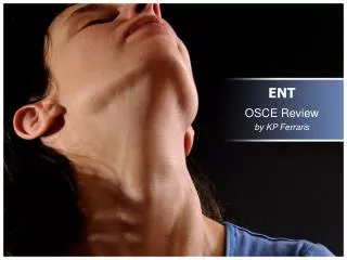
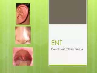
Eye and ENT Examination. Ear, Nose, and Throat Anatomy. Oral Cavity. Pharynx Uvula Tonsils. Lymph Nodes. 1 - Anterior cervical 2 - Posterior cervical 3 - Preauricular. 3. 1. 2. Ears. Pinna. Ear Examination. 3 steps Outer ear Canal Inner ear

Eye and ENT Examination
E N D
Presentation Transcript
Oral Cavity • Pharynx • Uvula • Tonsils
Lymph Nodes • 1 - Anterior cervical • 2 - Posterior cervical • 3 - Preauricular 3 1 2
Ears Pinna
Ear Examination • 3 steps • Outer ear • Canal • Inner ear • General inspection and palpation (outer ear) • Otoscopic examination (canal and inner ear)
Outer Ear Exam • Look for deformities, lumps, skin lesions (eg. Herpes lesions, hematomas) • Associated structures • Lymph nodes • Pharynx • Eustachian tube
Ear Canal and Inner Ear Exam • Use proper sized otoscope tip • Turn on otoscope and check that light works • Pull ear up, back, and toward you • Use pinky finger for support and to prevent injury • Place otoscope tip in ear canal, then lean forward and start looking into the otoscope
Ear Canal Exam • Look for redness, swelling, discharge, foreign bodies, wax • Pain with tragus and pinna manipulation can indicate problem with canal, as opposed to inner ear
Examination of TM • The TM is clear (transparent) when light passes through the membrane • The TM is dull (opaque) when light does not pass through the membrane so that the bony landmarks can not be clearly seen
Bulging TM • The bulging often impairs the visibility of the landmarks
Lymph Nodes • 1 - Anterior cervical(pharyngitis) • 2 - Posterior cervical(mono) • 3 – Preauricular(conjunctivitis) 3 1 2
Lymph Nodes • Roll the lymph node area under the pads of your fingers, compressing it against the underlying structures • Feel for size and tenderness • Check for symmetry • Is there an enlarged gland that just happens to be on the side of the earache or sinus pressure?
Sinuses • Grasp head fairly firmly, and push with thumbs on the frontal and maxillary areas • For ethmoid sinuses, squeeze firmly between eyes with thumb and index finger • Evaluate for tenderness • Fairly nonspecific
Eye Examination • 4 components • 1 - Visual acuity (Snellen eye chart) • 2 - Visual fields (by confrontation) • 3 - Extra ocular movements (“H”) • 4 - Ophthalmoscopic examination
1- Visual Acuity Exam • Use a Snellen eye chart • 20 feet from chart • Read at least half of each line • Read posters, magazines, newspaper if nothing else is available
2 - Visual Fields Exam(by Confrontation) • Have patient cover one eye lightly and look at your nose • Stand facing patient (confronting them) holding hands out to sides • Check upper fields by wiggling fingers of one or both hands • Repeat for lower fields • Repeat for other eye
3 - Eye Movements Exam(Extra ocular movements) • Ask patient to look at your fingers • Keep head still. Stabilize chin if needed • Make large “H” • Convergence test (bring finger to their nose) • Cross eyed
4 - Ophthalmoscopic Exam • Adjust proper settings on scope (light intensity, light shape, light color, focus) • Position patient and adjust ambient lighting • Start laterally from a distance and obtain red reflex, then approach steadily as if peering through a keyhole • If lots of glare, use a smaller diameter light setting • Find optic disc directly or by following a blood vessel from narrower aspect to wider aspect, which will lead you to the optic disc
Generally speaking, you don’t have to worry about rupturing a patient’s tympanic membrane with a typically placed otoscope tip because, a. The tympanic membrane is tough like shoe leather b. They still have another good ear anyway! c. The canal is about 1 inch deep c
Match the Swollen Lymph Nodes! Anterior cervical nodes onlyAnterior and posterior cervical nodesPreauricular nodes Possible Diagnoses – Herpes infection on temple, mono, tonsillitis Tonsillitis Mono Herpes
If you were to tug on someone’s pinna or poke someone’s tragus you would expect, a. A slap b. Pain which could indicate an acute otitis media c. Pain which could indicate otitis externa c
To look in a person's ear, you would move the pinna in the following direction. a. Down and out b. Up, back, and toward you c. Straight up d. Consult your GPS, then proceed as directed b
If you were looking in a healthy right ear, you would expect to see which of the following? a. The cone of light to the left b. Our new kitty from last year c. Same cat this year d. A shiny, somewhat transparent-appearing tympanic membrane d
Matching Game?If you wanted to evaluate someone’s visual acuity, you would (choose either best, pretty good, bad, or stupid!), Ignore their complaint of decreased vision altogether Use a Snellen eye chart Poke them in the eye with your ophthalmoscope Have them read from a magazine Bad Best Stupid Pretty good
Which of the following is the single greatest scientific achievement of all time? Einstein’s theory of relativity. The concept of the number “0”. Darwin’s discovery of Evolution. Vaccination. None of the above. It’s the Mr. Clean Magic Eraser.