Download
1 / 23
240 likes | 415 Vues
Cells of the Immune System. Dr. Raid Jastania. Intended Learning Outcomes:. Students should be able to describe the immune system and the cellular interactions involved in humoral and cellular immunity.

E N D
Cells of the Immune System Dr. Raid Jastania
Intended Learning Outcomes: • Students should be able to describe the immune system and the cellular interactions involved in humoral and cellular immunity. • Students should have a clear understanding of major histocompatibility complex (MHC) molecules and their role in immunity. • Students should be able to list the hypersensitivity reactions, and discuss the basic mechanism of them and give few examples of each type. • Students should understand how “Tolerance” occurs and how failure of tolerance may result in autoimmune diseases • Students should have a clear understanding of the pathogenesis of autoimmune diseases, and know the features of SLE. • Students should have a general brief understanding of immunodeficiency.
Diseases of the immune system: • There are 3 types or categories of diseases of the immune stystem: • Hypersensitivity reactions • Autoimmune diseases • Immunodeficiency
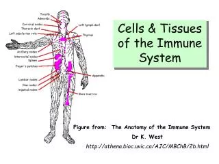
Definitions • Central immune system: bone marrow and thymus • Peripheral immune system: any site except bone marrow and thymus, it is mainly peripheral blood, lymph and lymph nodes, spleen and mucosa of GIT, respiratory system and genitourinary system. • Peripheral Lymphocytes: Lymphocytes not in the central part of the immune system (not in bone marrow or thymus) • Naïve cell: used to describe newly produced lymphocytes that have not encounter antigens.
Definitions • Transformed cell: changes of the morphology of lymphocytes when they encounter antigen and start the process of adaptive immunity. • Maturity and Differentiation and CD’s: The lymphocytes pass through different stages of maturation. During each stage, they express certain molecules on the cell surface (or in the cytoplasm). These molecules are named cluster of differentiation (CD). CD’s may appear at one stage then disappear at another.
Definitions • TCR: T-cell receptor is the molecule that T cells can recognize antigens with and start adaptive response. • BCR: B-cell receptor is IgM molecule that binds to antigens to stimulate B-cells. • APC: antigen presenting cell, including macrophages, monocytes, dendritc cells, and B-cells.
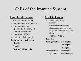
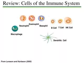
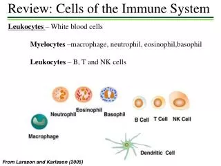
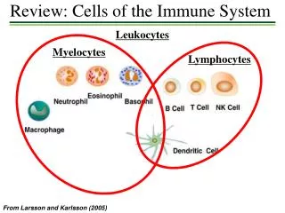
Cells of the Immune system • T-lymphocytes: • Thymus-derived • Mediate and control immunity • 60-70% of circulating lymphocytes • Found in the thymus, lymph nodes, spleen • TCR (T cell receptor) recognizes specific antigen • TCR needs somatic rearrangement to be unique for antigens
T-cells • TCR consists of alpha and beta chains, each has variable region and a constant region • There is gamma, delta TCR as well, less in number, found in mucosa • TCR is accompanied by CD3 (consists of alpha, delta, and epsilon chains) and 2 zeta chains. • CD4 and CD8 subsets: CD4+ is T helper cell. CD8+ is T cytotoxic cell.
T-cells • T helper is further divided to two subtypes: • T-h1: mediate cellular immunity by secreting IL-2, and IFN-gamma • T-h2: mediate humoral immunity by secreting IL-4, IL-5, IL-10
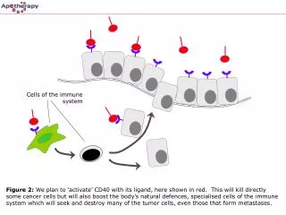
T-cells • The interaction of T-cells with APC’s needs 2 signals: • TCR binds to antigen, and CD4/8 binds to MHC-II/I • CD28 binds to B7-1 or B7-2 • In the absence of the second signal, T-cell either die or be inactivated (anergy)
B-lymphocytes • Bone marrow-derived • 10-20% of circulating lymphocytes • Found in lymph, and lymph nodes, spleen tonsils and all mucosal surfaces. • BCR is IgM molecule that binds to antigens • The final step of maturation is plasma cell which secrete antibodies. • There are 5 classes of anibodies: IgG, IgM, IgE, IgA, IgD • CD21 is the receptor for EBV.
B-lymphocytes • The Journey of B-cells
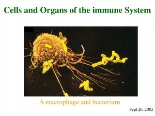
Macrophages • APC: antigen presenting cell. Processing and presentation • Express MHC-II that interact with CD4+ T-cells • Secrete cytokines • Kill microbes coated by antibodies and complement
Dendritic Cells • Interdigitating dendritic cells are found in lymphoid organs, interstitial tissue of visceral organs eg. Lung, skin. • Follicular dendritic cells: express Fc receptor that binds to Ag-Ab complex
Natural Killer Cells • 10-15% of peripheral lymphocytes • can lyse cells without previous sensitization • express Killer Inhibitor Receptor (KIR). When KIR binds to MHC-I it prevents killing • CD16 is Fc receptor • Mediate Antibody-dependent Cell-mediated Cytotoxicity
Major Histocompatibility Complex (MHC), Human Leukocyte Antigen (HLA) • MHC or HLA are molecules that are expressed on the cell membrane of cells. They are polymorphic and each person has a unique MHC. • They were first described in transplant rejection and called HLA • MHC restriction means that T-cells do not recognize antigens unless they are presented by APC with MHC.
MHC, HLA:There are 3 main classes • Class I: MHC-I: • are encoded in Chromosome 6 • HLA-A, HLA-B, HLA-C • The extracellular portion contains cleft for Ag presentation to CD8+ T-cells • Class I is present on all nucleated cells • Generally it presents proteins that have been synthesized in the cell eg. Viral proteins
Class II: MHC-II: HLA-D (HLA-DP, HLA-DQ, HLA-DR) The extracellular portion contain cleft for Ag binding and presentation to CD4+ T-cells Expressed on APC (monocytes, macrophages, dendritic cells) and B-cells. May be expressed on other cells if induced by IFN-gamma, eg. Endothelium, fibroblasts, renal tubular epithelium. It generally binds and present proteins that come from outside the cell, eg. Bacterial proteins
Class III: MHC-III: • Include some of the complement components C2, C3 and TNF • They are not Histocompatibility molecules
Diversity of MHC • Class I: 6 different molecules (genes) in a person • Class II: each molecule consists of alpha and beta chains, can give 20 different combination in a person. • Overall: innumerable combination of molecules that give a person unique MHC • HLA typing is done for organ transplantation • There is different ability of MHC’s (and persons) to bind and present antigens, and hence different immune response.
HLA and disease association • Many diseases are associated with selected HLA type • Example: ankylosing spondylitis associated with HLA-B27 with 90x increase risk of developing the disease • HLA is associated with 3 groups of diseases: • Inflammatory diseases, eg. Ankylosing spondyliti • Inherited errors of metablolism, eg. 21-hydroxylase deficiency (HLA-BW47) • Autoimmune diseases, eg. Rheumatoid arthritis
Cytokines • Low molecular weight polypeptides • Secreted by lymphocytes and monocytes • Less secreted by epithelial and mesenchymal cells • Act on receptors, eg. IL-2 acts on IL-2R • Act by autocrine, paracrine and endocrine fashion • Different action on different cells • Eg. IL-2 activate T-cells, and regulate B-cells • Some of them are antagonists, eg. IFN-gamma opposes IL-10
Classes of Cytokines • Cytokines that mediate innate immunity: eg. IL-1, TNF, IL-6 • Cytokines that regulate lymphocyte growth, activation and differentiation, eg. IL-2, IL-4, IL-5, IL-12, IL-15, TGF-alpha • Cytokines that activate inflammatory cells, eg. IFN-gamma, TNF • Chemokines: mediate chemotaxis, IL-8 attract neutrophils, IL-5 attract eosinophils • Cytokines that stimulate hematopoiesis, eg. GM-CSF granulocyte and macrophage colony stimulating factor