Tumor biology
460 likes | 1.19k Vues
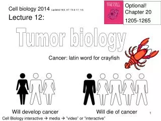
Optional! Chapter 20 1205-1265. Cell biology 2014 (updated 18/2, 4/7 - 13 & 1/1 -14). Lecture 12:. Tumor biology. Cancer: latin word for crayfish. Will develop cancer. Will die of cancer. Cell Biology interactive media ”video” or ” interactive ”. Basic tumor nomenclature.

Tumor biology
E N D
Presentation Transcript
Optional! Chapter 20 1205-1265 Cell biology 2014 (updated 18/2, 4/7 -13 & 1/1 -14) Lecture 12: Tumor biology Cancer: latin word for crayfish Will develop cancer Will die of cancer Cell Biologyinteractive media ”video” or ”interactive”
Basic tumor nomenclature Benign tumor Malignant tumor = cancer Metastasis forming cell (primary killer) Carcinoma: derived from epithelial cells (90% of all cancers) Sarcoma: derived from connective or muscle tissue Leukemia: derived from hematopoieticcells (BM and blood) Lymphoma: derived from lymphocytes (lymph nods)
Different views on cancer biology Old fasion view A heterotypic cell biology view Immune cells Non-autonomous heterogeneous cancer cells Autonomous cancer cells Other cells Endothelial cells In vitro propagated cell lines can only rarely be established from tumor biopsies tumor cells depends on their specific surrounding
Tumor progression A malignant tumor does not arise from a single genetic change; many changes are required to produce a life threatening cancer Tumor progression is defined as the acquisition of permanent changes in characteristics of selected subpopulations of the tumor Progenitors of the same clone, but still a heterogeneous tumor
CKI Ras Bcl-2 Rb Definitions: oncogenes and tumor suppressors An oncogene is a gene that when mutated, or overexpressed, contributes to converting a normal cell into a tumor cell (constitutive activity dominant phenotype) Onc point mutation overexpression A tumor suppressor-gene is a gene whose loss, or inactivation, contributes to converting a normal cell into a tumor cell (recessive phenotype) TS p53 Inactivating point mutations or loss of the entire gene (germ line mutation in one allele and/or acquired somatic mutations)
The normal stability of the genome makes cancer development statistically improbable Tumors acquire the capability to rapidly accumulate genetic changes by e.g., the following mechanisms: 1.Microsatellite INstability (MIN): Point mutations Common causes: defective DNA mismatch repair genes 2.Chromosomal INstability (CIN): Aneuploidy Common causes: aberrant centrosome numbers defective spindle regulatory proteins defective checkpoint control 3. Chromosome breaks and translocations Common causes: eroded telomeres DNA breaks
Common cause of gene loss and amplification DNA strand break DNA duplication End fusion Chromosome separation, novel breaks Gene loss TS DNA break Telomere Gene amplification Onc
The six hallmarks of cancer – A cell biology perspective 1. Self-sufficiency in proliferative signals 2. Insensitivity to anti-growth signals 3. Evading cell death (apoptosis) 4. Limitless replicative potential Make new blood vessel! 5. Sustained angiogenesis 6. Metastasis capability Adopted from Hanahan & Weinberg, Cell 2000
Tumor progression - molecular mechanisms • To be able to turn into a malignant tumor, each of the six • hallmarks has to be fulfilled • This is done by changing the level/activity of various proteins Only one protein per pathway needs to be changed! For example, a single protein in a mitogenic signaling pathway: G1 G1 myc myc Even if two tumors would belong to the same diagnostic group, they still have a unique combination of genetic alterations
P P Cdk Cdk G1 S DNA replisome 1. Self-sufficiency in proliferative signals Mitogenic signaling (growth promoting signals) Onc Onc Onc The retinoblastoma pathway TS Production of S cyclin Rb E2F Initiation of replication Cdc6 DNA replisome Mcm ORC Production of DNA replisome components
1. Cell type specific mitogenic pathways Cells from different tissues express distinct sets of growth factor receptors and signaling proteins Cell type B Cell type C Cell type A Major mitogen signaling pathway: RTK Wnt Hedgehog Alterations in tumors:RTK signaling Wnt signaling Hedgehog signaling
Wnt RTK Frizzled Ras APC 1. Aberrant proliferative signals in tumors XGF Hedgehog Onc Onc TS Patched Smoothened Onc Dishevelled TS TS Raf Axin GSK-3b Fused Onc SuFu Erk TS b-catenin Onc myc Onc myc G1 Gli Gli Onc Onc Onc Onc myc Onc Onc G1 G1
P P Cdk Cdk G1 S TGF-b DNA replisome 2. Insensitivity to anti-growth signals Mitogen signaling p15 p21 p16 TS TS viral The retinoblastoma pathway HPV E7 Onc Rb E2F Cdc6 ORC
p53 BH3 only TS TS Onc Bax Bcl-2 Cyt. C 3. Evading cell death (apoptosis) Ligand Survival factor signaling Onc Death receptor TS Adaptor Caspase 8 Caspase 9 Caspase 3 Apoptosis
P P P P P 3.Survival factor signaling PI-3 K Onc or GPCR or RTK 3 3 PTEN Onc TS Onc PKB/Akt elF4E Bad G1 Cell growth + Apoptosis
A G G G T T 4. Limitless replicative potential Telomeres: stretches of repetitive DNA at the chromosome ends that can form a protective loop structure Chromosome lacking telomeres will trigger a p53 dependent cell cycle block 5´ 5´ 3´ 3´ Complementarity due to the repetitive sequence 5´ -GGGTTAGGGTTAGGGTTA Telomerase, usually not expressed in somatic cells AUCCCAAU 3´ -CCCAATCC To maintain telomere length tumor cells can re-start expression of telomerase. An alternative mechanism employs enzymes that are involved in DNA recombination
Make new blood vessel! 5. Sustained angiogenesis Blood vessel < 100 mm Endothelial cell Diffusion of O2 and nutrients Too long I am suffocating! Let’s express VEGF VEGF: Vascular endothelial growth factor Anim. 23.7-angiogenesis
Ras Ub Ub Ub 3. 2. 1. 4. 5. Vascular Endothelial Growth Factor - VEGF Ras dependent signaling can increase expression levels of HIF-1 Onc Constitutively produced in all tissues HIF-1 VEGF VEGF gene Constitutively degraded via pVHL, unless… pVHL HIF-1 TS …hypoxia (low O2) HIF-1: Hypoxia induced factor pVHL: von Hippel–Lindau syndrome (hereditary cancer) is caused by a germline mutation in the VHL gene Proteosome
p53 p53 Loss of p53 loss of angiogenisis inhibition 5. Angiogenic factors affecting endothelial cells Activators Inhibitors VEGF Thrombospondin-1 Tumor with active p53 Tumor without active p53 No angiogenesis Angiogenesis
Ras Avastinä 5. Summary: regulation of angiogenesis Onc p53 HIF-1 TS pVHL TS Thrombo- spondin-1 VEGF Angiogenesis
40-120 nm 1. 2. 1. 2. 3. 4. 4. 6. Metastasis capability Metastasis, the ability of cancer cells to migrate, results from multiple mutation events Basal lamina 3. Loss of cell-cell adhesion Loss of hemidesmosomes Proteolytic degradation of the ECM Migration through the ECM
6. Example of loss of cell-cell adhesion Loss of E-cadherin is an important step in generating daughter tumors (metastasis) in carcinomas Malignant tumor Benign tumor Tumor progression TS Migration, resettlement and further proliferation • Loss of E-cadherin • decreased cell adhesion Metastasis
1. 2. 3. 1. 2. 3. 6. Penetration of basal lamina Collagen IV fibril Laminin Reprogramming / de-differentiation of cells: Loss of hemidesmosomes/lamininreceptor (integrin) Expression of collagenase Cytoskeletal changes Epithelial–mesenchymal transition (EMT)
6. Making it through the connective tissue Cell secretes proteases to clear a path through the ECM Blood vessel
6. Sites of metastasis – blood flow • Blood flow pattern determine the metastasis pattern in most • case (~70%) Capillary of the lung Tumor cell entering the blood system Lung metastasis Capillary of the liver Stomach or intestinal tumor cell entering the blood system Liver metastasis
6. Sites of metastasis – microenvironment • ”Seed-soil” pattern determine the metastasis pattern in other • cases (~30%) Capillary of the lung Prostate tumor cell entering the blood system No lung metastasis due to non- favorable ”climate” Adjacent bone cells produce specific factors needed for tumor cell growth X X Capillary of a bone
X X X X X X X X X X X X 6. 5. 1. 2. 4. 4. 3. 5. 1. 2. 3. 6. Tumor progression: Familial adenomatous polyposis Alberts et al. Fig. 20-46 Self-sufficiency in proliferative signals Insensitivity to Anti-growth signals Limitless replicative potential Sustained angiogenesis Evading cell death Metastasis capability
Wnt APC APC APC Chromosomal instability Self-sufficiency in proliferative signals Metastasis capability Step I.Starting point of familial adenomatous polyposis By chance loss of the intact APC allele! TS b-catenin Axin GSK-3b MMP7 G1 Note dual action of APC: Hallmarks 1 & 6
- Ras p15 TGF b Step II. Progression of colon carcinoma Loss of SMAD4 Oncogenic mutation in RAS XGF Smad 4 TS Onc VEGF G1 Sustained angiogenesis Self-sufficiency in proliferative signals Insensitivity to anti-growth signals Hallmarks 1, 2 & 5
Bax Bcl-2 p21 Step III. Progression of colon carcinoma Loss of p53 DNA damage p53 Aberrant/incomplete proliferation signals TS Thrombo- spondin-1 PUMA Insensitivity to anti-growth signals Sustained angiogenesis Evading cell death Hallmarks 2, 3 & 5
Step IV. Progression of colon carcinoma Loss of E-CADHERIN Expression of telomerase AUCCCAAU TS Limitless replicative potential Metastasis capability The End Hallmarks 4 & 6
Ras Ras APC APC Fulfilling the hallmarks of cancer in colon cancer 1. Self-sufficiency in proliferative signals p53 2. Insensitivity to anti-growth signals Smad 4 p53 3. Evading cell death (apoptosis) Telomerase 4. Limitless replicative potential AUCCCAAU 5. Sustained angiogenesis p53 6. Metastasis capability E cadherin Fulfilling hallmarks 1 – 6 within a life time depends on genomic instability
Breast cancer in Sweden • 6,623 new cases in 2002 (earlyonset) • Incidence/year~115 per 100,000 • Mortality/year~ 35 per 100,000
Tumor/ Node/ Metastasis: T, clinical/mammographic evaluation of tumor (0-4). N, clinical evaluation of regional lymph nodes (0-3). M, distant metastases (0, 1). Stage 0: Tis N0 M0 Stage I: T1 N0 M0 Stage 2: T1-3 N1 M0 Stage 3: T1-4 N1-3 M0 Stage 4: T1-4 N1-3 M1 (is: in situ well encapsulated) The TNM system for clinical staging
Routine prognostic and predictive factors • TNM (Tumor/Node/Metastasis) • Histologic type and grade (as judge by the appearance under the microscope) • Molecular markers: Ki-67, estrogen and progesteron receptors, and ERBB2 (EGF receptor).
Decision tree: breast cancer treatment at NUS T: clinical/mammographic evaluation N: regional lymph nodes M: distant metastases Non-specific Mix of cytostatic drugs, e.g., FEC (5-FU, epirubicin, cyklofosfamid) or SBG 2000-1 mix Irradiation therapy Specific TAM: Tamoxifen (anti-estrogen) A temporary cure! (3-60 (?) years)
Future goals of (molecular) diagnostics • Early detection • Accurate prognosis • Good prediction (of therapy response) • Reveal molecular therapy targets