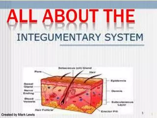
ALL ABOUT THE SPINAL CHORD
410 likes | 1.19k Vues
ALL ABOUT THE SPINAL CHORD. Functions of Spinal Cord. Final common pathway for the somatomotor system Conveys somatosensory information from the body Autonomic neurons Reflexes Central pattern generators for rhythmic movements. Sensory vs motor roots.

ALL ABOUT THE SPINAL CHORD
E N D
Presentation Transcript
Functions of Spinal Cord • Final common pathway for the somatomotor system • Conveys somatosensory information from the body • Autonomic neurons • Reflexes • Central pattern generators for rhythmic movements
Sensory vs motor roots Ventral: somatic motor + visceral motor
Motor Neurons • Alpha and gamma motor neurons • aka ventral horn cells, anterior horn cells • Very elaborate dendritic tree • Neurotransmitter=Ach • Alpha: extrafusal fibers • Gamma: intrafusal fibers
Spinal Cord and Spinal Nerves • Spinal cord • Spinal nerves
Overview of Spinal Cord • Information highway between brain and body • Extends through vertebral canal from foramen magnum to L1 • Each pair of spinal nerves receives sensory information and issues motor signals to muscles and glands • Spinal cord is a component of the Central Nervous System while the spinal nerves are part of the Peripheral Nervous System
Functions of the Spinal Cord • Conduction • bundles of fibers passing information up and down spinal cord • Locomotion • repetitive, coordinated actions of several muscle groups • central pattern generators are pools of neurons providing control of flexors and extensors (walking) • Reflexes • involuntary, stereotyped responses to stimuli (remove hand from hot stove) • involves brain, spinal cord and peripheral nerves
Anatomy of the Spinal Cord • Cylinder of nerve tissue within the vertebral canal (thick as a finger) • vertebral column grows faster so in an adult the spinal cord only extends to L1 • 31 pairs of spinal nerves arise from cervical, thoracic, lumbar and sacral regions of the cord • each cord segment gives rise to a pair of spinal nerves • Cervical and lumbar enlargements • Medullary cone (conus medullaris) = tapered tip of cord • Cauda equinae is L2 to S5 nerve roots resemble horse’s tail
Meninges of the Spinal Cord • 3 Fibrous layers enclosing spinal cord • Dura mater • tough collagenous membrane surrounded by epidural space filled with fat and blood vessels • epidural anesthesia utilized during childbirth • Arachnoid mater • layer of simple squamous epithelium lining dura mater and loose mesh of fibers filled with CSF(creates subarachnoid space) • Pia mater • delicate membrane adherent to spinal cord • filium terminale and denticulate ligaments anchor the cord
Cross-Sectional Anatomy of the Spinal Cord • Central area of gray matter shaped like a butterfly and surrounded by white matter in 3 columns • Gray matter = neuron cell bodies with little myelin • White matter = myelinated axons
Gray Matter in the Spinal Cord • Pair of dorsal or posterior horns • dorsal root of spinal nerve is totally sensory fibers • Pair of ventral or anterior horns • ventral root of spinal nerve is totally motor fibers • Connected by gray commissure punctured by a central canal continuous above with 4th ventricle
White Matter in the Spinal Cord • White column = bundles of myelinated axons that carry signals up and down to and from brainstem • 3 pairs of columns or funiculi • dorsal, lateral, and anterior columns • Each column is filled with named tracts or fasciculi (fibers with a similar origin, destination and function)
Spinal Tracts • Ascending and descending tract head up or down while decussation means that the fibers cross sides • Contralateral means origin and destination are on opposite sides while ipsilateral means on same side
Organization of Somatosensory System • Touch • Fine, discriminitive touch • Presssure and vibration sense • Conscious proprioception • Unconscious proprioception • Pain • Temperature http://thalamus.wustl.edu/course/body.html
Dorsal Column Ascending Pathway • Deep touch, visceral pain, vibration, and proprioception • Fasciculus gracilis and cuneatus carry signals from arm and leg • Decussation of 2nd order neuron in medulla • 3rd order neuron in thalamus carries signal to cerebral cortex
Spinothalamic Pathway • Pain, pressure, temperature, light touch, tickle and itch • Decussation of the second order neuron occurs in spinal cord • Third order neurons arise in thalamus and continue to cerebral cortex
Spinoreticular Tract • Pain signals from tissue injury • Decussate in spinal cord and ascend with spinothalamic fibers • End in reticular formation (medulla and pons) • 3rd and 4th order neurons continue to thalamus and cerebral cortex
Spinocerebellar Pathway • Proprioceptive signals from limbs and trunk travel up to the cerebellum • Second order nerves ascend in ipsilateral lateral column
Corticospinal Tract • Precise, coordinated limb movements • Two neuron pathway • upper motor neuron in cerebral cortex • lower motor neuron in spinal cord • Decussation in medulla
Descending Motor Tracts • Tectospinal tract (tectum of midbrain) • reflex turning of head in response to sights and sounds • Reticulospinal tract (reticular formation) • controls limb movements important to maintain posture and balance • Vestibulospinal tract (brainstem nuclei) • postural muscle activity in response to inner ear signals
Poliomyelitis and ALS • Diseases causing destruction of motor neurons and skeletal muscle atrophy • Poliomyelitis caused by poliovirus spread by fecally contaminated water • weakness progresses to paralysis and respiratory arrest • Amyotrophic lateral sclerosis • sclerosis of spinal cord due to astrocyte failure to reabsorb glutamate neurotransmitter • paralysis and muscle atrophy
Anatomy of a Nerve • A nerve is a bundle of nerve fibers (axons) • Epineurium covers nerves, perineurium surrounds a fascicle and endoneurium separates individual nerve fibers • Blood vessels penetrate only to the perineurium
Anatomy of Ganglia in the PNS • Cluster of neuron cell bodies in nerve in PNS • Dorsal root ganglion is sensory cell bodies • fibers pass through without synapsing
The Spinal Nerves • 31 pairs of spinal nerves (1st cervical above C1) • mixed nerves exiting at intervertebral foramen • Proximal branches • dorsal root is sensory input to spinal cord • ventral root is motor output of spinal cord • Distal branches • dorsal ramus supplies dorsal body muscle and skin • ventral ramus to ventral skin and muscles and limbs • meningeal branch to meninges, vertebrae and ligaments
Branches of a Spinal Nerve • Each has dorsal and ventral ramus.
Rami of Spinal Nerves • Notice the branching and merging of nerves in this example of a plexus
Shingles • Skin eruptions along path of nerve • Varicella-zoster virus (chicken pox) remains for life in dorsal root ganglia • Occurs after age 50 if immune system is compromised • No special treatment
Nerve Plexuses • Ventral rami branch and anastomose repeatedly to form 5 nerve plexuses • cervical in the neck, C1 to C5 • supplies neck and phrenic nerve to the diaphragm • brachial in the armpit, C5 to T1 • supplies upper limb and some of shoulder and neck • lumbar in the low back, L1 to L4 • supplies abdominal wall, anterior thigh and genitalia • sacral in the pelvis, L4, L5 and S1 to S4 • supplies remainder of lower trunk and lower limb • coccygeal, S4, S5 and C0
Some Facts • Human Nervous System: • 31 pairs of spinal nerves • Divided into cervical, thoracic, lumbar and sacral levels • Spinal nerves impose segmentation; actual cord consists of columns of cells • Innervation of body is segmented (dermatomes)
Cutaneous Innervation and Dermatomes • Each spinal nerve receive sensory input from a specific area of skin called dermatome • Overlap at edges by 50% • a total loss of sensation requires anesthesia of 3 successive spinal nerves