Hematopathology Lab
Hematopathology Lab. December 12, 2013. Case 1. Normal Peripheral Blood Smear. Q1. Describe the normal morphology of the red blood cells (RBCs) Q2. Identify and describe the white blood cells (WBCs) in the smear

Hematopathology Lab
E N D
Presentation Transcript
Hematopathology Lab December 12, 2013
. Normal Peripheral Blood Smear
Q1. Describe the normal morphology of the red blood cells (RBCs) Q2. Identify and describe the white blood cells (WBCs) in the smear Q3. Identify the platelets in the smear and describe their characteristics. How many do you see in the high power field?
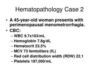
History A 45-year-old woman presents with perimenopausal menometrorrhagia. CBC: WBC 9.7x10³/uL Hemoglobin 7.8g/dL Hematocrit 23.5% MCV 73 femtoliters (fL) Red cell distribution width (RDW) 22.1 Platelets 187,000/uL
-RBCs are smaller than Patient’s Blood Smear Normal
Q1 Describe the morphology of the RBCs. Contrast to the normal peripheral blood smear. Q2 Correlate the findings on the peripheral smear with the CBC indices.
Q3 What is your diagnosis? Q4 What is the etiology in this patient? What are other etiologies?
Q5 What symptoms and physical exam findings may be associated with this condition? Q6 What is “Plummer Vinson Syndrome”?
Case History A 22-year-old African-American man presents with severe pain in several joints and diffuse abdominal pain. He states he is active physically and participates in different sports several times a week. He has had no fevers or chills. Review of systems is negative for any symptoms of infection.
CBC findings: WBC 13.2x10³/uL Hemoglobin 7.9g/dL Hematocrit 22.8% MCV 91.4 femtoliters (fL) RDW 24.1 Platelets 481,000/uL
Q1 Describe the morphology of the RBCs. Q2 What is your diagnosis? Q3. Define “poikilocytosis” and how it is demonstrated on the smear.
Q4. What are the most common genetic mutations associated with this disease? Q5. Explain the etiology of the patient’s symptoms. Why was the absence of symptoms of infection specifically noted?
Q6 Describe the likely gross morphology of this patient’s spleen. What are the clinical implications? Q7 Define “acute chest syndrome”.
CASE 4 History A 60 year-old man presents with mild fatigue. Further questioning reveals a vague feeling of abdominal “fullness” and more bruising of his skin. Physical examination is remarkable for splenomegaly to the level of the umbilicus.
CBC findings: WBC 75.1x10³/uL Hemoglobin 8.5g/dL Hematocrit 25.5% Platelets 56,000/uL MCV 88.4 femtoliters (fL) RDW 16.1 The hematology analyzer has flagged the specimen for possible immature WBC forms
-WBCs are markedly increased in number, predominantly cells of the neutrophil series. -Many mature neutrophils are seen as well as earlier forms showing a)less nuclear segmentation (bands, myelocytes) and b) prominent primary granules without secondary fine pink granulation (promyelocytes). No blasts are seen. Peripheral Blood SmearImage A
Q1 Describe the findings on the peripheral smear. Do you agree that there is an increased number of immature WBC forms in the peripheral blood? Name the cells indicated by the arrows.
Q2 There was also an increased number of cells highlighted by the arrow in peripheral blood smear image B. What is the cell?
Q3 What do “A” and “B” represent? Q4 On low power, what is the most striking difference between the patient’s bone marrow and the normal bone marrow?
Q5 What is your diagnosis? Q6 Define “leukemoid reaction”. What distinguishes leukemoid reaction from our patient’s diagnosis?
Q7 How do the history and physical examination findings relate to the diagnosis and to the CBC results?
Q8 The image is from a patient with the same disease who underwent autopsy examination. Explain the findings.
Q9 What is the clinical significance of the identified findings on the karyotype?
Reactive Lymph NodeLow Power At low magnification, the lymph node is surrounded by a thin capsule o B A Q1 Describe the architecture. Identify/describe the structures highlighted by arrows, circle
Higher Power On higher magnification, germinal centers show normal spacing in between in each other Q2 Name the structures highlighted by the asterix. What is “A”? * A *
Q3 What types of cells compose the structure highlighted by the asterix? High Power *
Q4 What size are normal lymph nodes? Q5 Are peripheral lymph nodes palpable in normal healthy individuals?
History A 35 year-old man presents for evaluation of an enlarged non-tender cervical lymph node. The node has shown progressive enlargement over the past 4 months. The patient denies other symptoms such as fevers, night sweats, weight loss or fatigue.
Q1 Develop a differential diagnosis (using broad categories) for cervical lymphadenopathy
CBC WBC 7.2x10³/uL Hemoglobin 14.2g/dL Hematocrit 43.3% MCV 87.0 femtoliters (fL) RDW 14.1 Platelets 372,000/uL An excisional biopsy of a enlarged lymph node is performed.
Bisected lymph node Q2 Comment on the size of the lymph node. Describe the gross findings.
Low power Q3 Describe the histologic findings. Compare to the reactive lymph node. What is the asterix highlighting? * Reactive Node
Within the nodules, Q4 Describe the higher power findings.
Q5 Describe the high power findings. What is the arrow highlighting?
Q6 What is your diagnosis? Q7 What are other common histologic subtypes of this disease? Q8 What does the term “B Symptoms” refer to?
History A 51 year-old man presents with fatigue, malaise and occasional low grade fevers. Physical examination reveals bilateral non-tender cervical lymphadenopathy and an enlarged inguinal lymph node.