Download
1 / 1
10 likes | 169 Vues
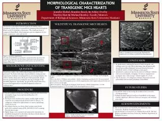
MORPHOLOGICAL CHARACTERIZATION OF TRANSGENIC MICE HEARTS. 0. Jennifer Heibel, Brandon Boeck , & Ashley Overby Marilyn Hart & Michael Bentley, Faculty Mentors Department of Biological Sciences, Minnesota State University-Mankato. INTRODUCTION. BACKGROUND AND SCIENTIFIC QUESTION.

E N D
MORPHOLOGICAL CHARACTERIZATION OF TRANSGENIC MICE HEARTS 0 Jennifer Heibel, Brandon Boeck, & Ashley Overby Marilyn Hart & Michael Bentley, Faculty Mentors Department of Biological Sciences, Minnesota State University-Mankato INTRODUCTION BACKGROUND AND SCIENTIFIC QUESTION The heart is a specialized muscle found in all animals with a circulatory system that is responsible for pumping blood throughout blood vessels by rhythmic contractions. The vertebrate heart is composed of myofibrils which maintain a precise alignment of two major components, actin and myosin. PROCEDURE WILDTYPE VS. TRANSGENIC MICE HEARTS FUTURE STUDIES ACKNOWLEDGEMENTS CONCLUSION Figure 3. Display of primary focus area of myofibril analysis – the inner heart chamber. F Figure 1. Sarcomere organization. The ends of actin filaments are attached to a structural anchor, the Z line, that maintains the alignment of the thin filament. Biochemical and cell biological studies suggest that actin capping protein (CP) attaches one end of the actin filament to the Z line. 1. Expand the study by increasing the number of mice tested in varying age groups. 2. Perform detailed statistical analysis of myofibrils to determine whether significant difference between transgenic and wildtype mice exists. • By qualitatively studying the hearts of both transgenic and wildtype mice we were able to determine that the genetically altered myocardium displayed increased disarray between myofibrils relative to their wildtype counterparts. • Quantitative analysis of mice hearts showed wildtypet-tubule’s with increased lengths, averaging 2.43 nm, in comparison to transgenic mice with average t-tubule lengths of 2.23 nm. • The 1% elastase / 1% collagenase solution effectively removed pericardium and revealed periodicity of myofibrils. In previous studies, mice with reduced expression of CP were generated. The hearts had major structural defects in muscle unit organization, leading to an enlarged heart and ensuing lethality. We wish to further characterize the hearts of the genetically altered mice by quantitatively measuring the spacing and length of the myofibril alignment. Specifically, do genetically altered mice have increased disorganization of myofibril alignment relative to wildtype mice? Figure 2. Qualitative comparison of myofibril alignment between wildtype and transgenic mice. Row one shows: Wildtype myocardium t-tubules displaying a periodicity of 2.43 nm (standard deviation of 0.11) at 850x magnification. A 2000x magnification of the region indicated by the red box is shown on the right. Row two shows:The display of transgenic heart muscle t-tubule arrangements, 2.23 nm (standard deviation of 0.11) in length, with same magnification as above. Figure 4. Scanning electron microscopy displaying low magnification of wildtype myofibril branching alignment. F 1. The myocardium of both the wildtype and genetically altered mice, six months of age, were removed and fixed in 2.5% glutaraldehyde. 2. The hearts were cut down the septum, and the right and left ventricles were independently treated with 1% elastase / 1% collagenase solution for eight minutes to remove underlying connective tissue. 3. The prepared tissue was freeze dried, sputter coated with gold, and visualized using a JOEL JSM 6510LV/LGS scanning electron microscope. 4. Digital images were captured and analyzed for alterations in myofibril arrangement to determine the basis of heart defect of both wildtype and transgenic mice. 1. This research project was funded by the Minnesota State University, Mankato Foundation Grant. 2. Special thanks to our mentors, Dr. Marilyn Hart and Dr. Michael Bentley and Minnesota State University, Mankato Department of Biological Sciences.