Download
1 / 35
350 likes | 473 Vues
This study explores genome segmentation through the application of quantitative cellular descriptors derived from large-scale RNAi perturbations. We detail an overview of the microscopy screening process and computational analysis, including metrics optimization and cell cycle analysis using Acumen technology. We assess DNA damage foci formation and classify genetic phenoprints through advanced image analysis workflows. The methodology includes retesting and validation of phenotypic responses, identifying gene clusters linked to biological processes and regulatory pathways.

E N D
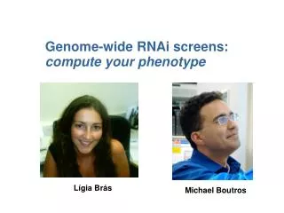
Segmenting the genome by quantitative cellular descriptors using large-scale RNAi perturbations Florian Fuchs, Oleg Sklyar, Gregoire Pau, Christoph Budjan, Thomas Horn, Wolfgang Huber and Michael Boutros
Contents • Overview microscopy screen • Computational analysis + metrics optimization (Greg) • Microscopy retest (with Qiagen library, BD pathway) • Cell cycle analysis with drug treatment (Acumen) • DNA damage foci formation (Zeiss AxioImager)
Metric optimization using STRING (Greg) score=quantile(d(i,j in STRING),0.1)/quantile(d(i,j in RANDOM),0.1
Metric optimization STRING: 59821 interaction pairs RANDOM: randomized version of STRING (nodes permutation)
Replicate analysis Pseudo-replicate siRNA comparison
Functional map edge if dist. < 0.1 38 clusters 1558 wells 32139 edges cyan – pos. control
Enrichment – hand-defined clusters • Find enrichment of GO and/or KEGG categories in a given cluster • Requirement: min. GO/KEGG terms size of 2, min. number of 2 genes per cluster belonging to the GO/KEGG category, p-value cut-off 1e-2 • 22 out of 38 clusters show enrichment in at least 1 GO category • e.g. chemotaxis, mitochondrion, regulation of cyclin-dependent protein kinase, cell cycle.
Enrichment – clusters defined by gene centers • Clusters based on a set of 29 genes of interest • e.g. CEP164, DONSON, CASP8AP2, CD3EAP, C20ORF4 • Radius of 30 closest genes to the gene of interest • Look for GO/KEGG enrichment in the 30 closest genes to these centers • Enrichment in e.g. centrosome, anti-apoptosis, mitosis, cell-cycle, proteasome
Donson - gene centered cluster 14 genes conserved in fly and worm
Donson gene centered cluster 30 closest genes to the center colored genes: GO enrichment
Microscopy retest Phenotypic readout (microscopy) with HeLa and U2OS • Individual single siRNAs (deconvolution of pools) • Pools from Qiagen (608) • Individual siRNAs from Ambion
Retest analysis of morphology screen Dharmacon retest Upgrade Validation on Phenotypic Level (56 Pools from Dharmacon) 4 % 13 % 24 % 20 % 39 % • Qiagen retest • 608 Genes were retested • 117 showed phenotype from global screen • additional 160 showed similar phenotype • Ambion single siRNA retest • 1 out of 2 plates finished • currently being analysed
Cell cycle analysis using Acumen Explorer Acumen eX3 • microplate cytometer for high-throughput in situ screening • applicable to a broad range of assays, e.g. • Cell cycle • Protein translocation • Cell proliferation • …
Cell cycle analysis using Acumen Explorer Plate formats: 96 to 1536 well plates in a high-throughput manner (25’ per 384 well plate) Laser: Up to 3 lasers – 405 nm (Hoechst, Alexa 405), 288 nm (PI, Alexa 488, FITC, eGFP), 633 nm (Alexa 633, Cy5) Features: Resolution similar to that achieved using a 20x microscope Field of view: 20x20mm (4 wells of a 96-well plate) => whole well scanning possible Multiplexing (up to 12 data channels)
Cell cycle analysis using Acumen Explorer Aim Identify genes involved in DNA damage signaling and/or are required for cell cycle progression Approach Downregulation of putative cell cycle regulatory proteins and drug treatment for induction of cell cycle arrest
Comparison positive and negative controls HeLa (750 c/w seeded) 24 hr post-transfection siRluc 1500 cells/well G1: 59%, S: 10%, G2/M: 29% [Frequency] 5 10^4 10^5 1.5 10^5 2 10^5 [Intensity] HeLa (750 c/w seeded) 24 hr post-transfection siPlk1 650 cells/well G1: 11%, S: 7%, G2/M: 80% [Frequency] 5 10^4 10^5 1.5 10^5 2 10^5 [Intensity]
Well view after classification of nuclear content Classification of cells into G1, S and G2/M phase by determining the DNA content of the cell G1 S G2/M
The pseudo time-lapse experiments are reproducible 720 genes 3 replicates siRNA incubation for 24, 36, 48 hr Red dots: controls
HU treatment causes cell cycle arrest [Frequency] [Intensity]
Results of HU treatment (time-lapse) 256 genes 48 hr siRNA +/- 24 hr HU + 0, 8, 16 or 24 hr release HeLa cell viablity phenotype after HU treatment
Results of HU treatment (time-lapse) 0 20 40 60 80 100 - HU
Results of HU treatment (time-lapse) 0 20 40 60 80 100 + HU
Microscopic analysis of DNA damage induction after siRNA treatment • Silencing of 24 target genes • 72 hr siRNA • 48 hr siRNA + 24 hr HU • 48 hr siRNA + 24 hr HU + 8 hr release • Staining of nucleus (Hoechst) and DNA-damage foci (anti-pH2AX) • Rluc, Top3a, Chek2, Clspn, Chek1, Casp8AP2, ERCC1, CDCA8, TMEM61, DLL4, Donson, ATM, CD3EAP, ATR, C20ORF4, PRIC285, SON, Rad17, RRMI, CADMI, Prim2A, WISPI, WDR33, Cep164
DNA damage pH2AX foci formation after 72 hr siRNA treatment (Zeiss AxioImager & Apotome, 63x-oil, deconvolution) DNA (Hoechst) α-pH2AX Rluc C20ORF4 Donson Donson
Donson depletion induces strong DNA damage foci formation (72 hr siRNA) DNA (Hoechst) α-pH2AX
CADM1 (cell adhesion molecule 1) May be involved in apoptosis, cell adhesion and regulation of cell cycle pH2AX-positive after release DNA (Hoechst) α-pH2AX 72 hr siRNA 48 hr siRNA + 24 hr HU + 8 hr release 48 hr siRNA + 24 hr HU
Outlook • Cloning of HA-tagged human Donson in pcDNA3.1 (o/e) • Generation of eGFP-tagged Donson fusion protein (pEGFP-C1), colocalization studies with known DNA damage markers • qPCR of single siRNAs of Donson (and others) • Cloning of mouse homolog of Donson in overexpression vector, rescue of Donson knockdown in human cells • Knockdown of fly homologs of Donson cluster in Drosophila cells, microscopy and cell-cycle (Acumen) analysis • Cell cycle analysis (Acumen) after IR irradiation & HU treatment in U2OS cells • Repeat DNA damage foci microscopy (replicate)