Download
1 / 110
1.17k likes | 2.19k Vues
Lecture 6: Osseous Tissue and Bone Structure. Topics:. Skeletal cartilage Structure and function of bone tissues Types of bone cells Structures of the two main bone tissues Bone membranes Bone formation Minerals, recycling, and remodeling Hormones and nutrition Fracture repair

E N D
Topics: • Skeletal cartilage • Structure and function of bone tissues • Types of bone cells • Structures of the two main bone tissues • Bone membranes • Bone formation • Minerals, recycling, and remodeling • Hormones and nutrition • Fracture repair • The effects of aging
The Skeletal System • Skeletal system includes: • bones of the skeleton • cartilages, ligaments, and connective tissues
Skeletal Cartilage • Contains no blood vessels or nerves • Surrounded by the perichondrium (dense irregular connective tissue) that resists outward expansion • Three types – hyaline, elastic, and fibrocartilage
Hyaline Cartilage • Provides support, flexibility, and resilience • Is the most abundant skeletal cartilage • Is present in these cartilages: • Articular – covers the ends of long bones • Costal – connects the ribs to the sternum • Respiratory – makes up larynx, reinforces air passages • Nasal – supports the nose
Elastic Cartilage • Similar to hyaline cartilage, but contains elastic fibers • Found in the external ear and the epiglottis
Fibrocartilage • Highly compressed with great tensile strength • Contains collagen fibers • Found in menisci of the knee and in intervertebral discs
Growth of Cartilage • Appositional – cells in the perichondrium secrete matrix against the external face of existing cartilage • Interstitial – lacunae-bound chondrocytes inside the cartilage divide and secrete new matrix, expanding the cartilage from within • Calcification of cartilage occurs • During normal bone growth • During old age
Bones and Cartilages of the Human Body Figure 6.1
Functions of the Skeletal System • Support • Storage of minerals (calcium) • Storage of lipids (yellow marrow) • Blood cell production (red marrow) • Protection • Leverage (force of motion)
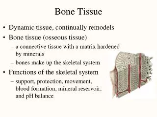
Bone (Osseous) Tissue • Supportive connective tissue • Very dense • Contains specialized cells • Produces solid matrix of calcium salt deposits and collagen fibers
Characteristics of Bone Tissue • Dense matrix, containing: • deposits of calcium salts • osteocytes within lacunae organized around blood vessels • Canaliculi: • form pathways for blood vessels • exchange nutrients and wastes
Characteristics of Bone Tissue • Periosteum: • covers outer surfaces of bones • consist of outer fibrous and inner cellular layers • Contains osteblasts responsible for bone growth in thickness • Endosteum • Covers inner surfaces of bones
Bone Matrix • Solid ground is made of mineral crystals • 2/3 of bone matrix is calcium phosphate, Ca3(PO4)2: • reacts with calcium hydroxide, Ca(OH)2 to form crystals of hydroxyapatite, Ca10(PO4)6(OH)2 which incorporates other calcium salts and ions
Bone Matrix • Matrix Proteins: • 1/3 of bone matrix is protein fibers (collagen) • Question: why aren’t bones made of ALL collagen if it’s so strong?
Bone Matrix • Mineral salts make bone rigid and compression resistant but would be prone to shattering • Collagen fibers add extra tensile strength but mostly add tortional flexibilitytoresist shattering
Chemical Composition of Bone: Organic • Cells: • Osteoblasts – bone-forming cells • Osteocytes – mature bone cells • Osteoprogenitor cells – grandfather cells • Osteoclasts – large cells that resorb or break down bone matrix • Osteoid – unmineralized bone matrix composed of proteoglycans, glycoproteins, and collagen; becomes calcified later
There are four major types of cells periosteum + endo endosteum only in matrix only
1. Osteoblasts • Immature bone cells that secrete matrix compounds (osteogenesis) • Eventually become surrounded by calcified bone and then they become osteocytes Figure 6–3 (2 of 4)
2.Osteocytes • Mature bone cells that maintain the bone matrix Figure 6–3 (1 of 4)
Osteocytes • Live in lacunae • Found between layers (lamellae) of matrix • Connected by cytoplasmic extensions through canaliculi in lamellae (gap junctions) • Do not divide (remember G0?) • Maintain protein and mineral content of matrix • Help repair damaged bone
3. Osteoprogenitor Cells • Mesenchyme stem cells that divide to produce osteoblasts • Are located in inner, cellular layer of periosteum • Assist in fracture repair
4. Osteoclasts • Secrete acids and protein-digesting enzymes Figure 6–3 (4 of 4)
Osteoclasts • Giant, mutlinucleate cells • Dissolve bone matrix and release stored minerals (osteolysis) • Often found lining in endosteum lining the marrow cavity • Are derived from stem cells that produce macrophages
Homeostasis • Bone building (by osteocytes and -blasts) and bone recycling (by osteoclasts) must balance: • more breakdown than building, bones become weak • exercise causes osteocytes to build bone
Osteoprogenitor cells osteoblasts osteocytes Osteoclasts are related to macrophages (blood cell derived) Bone cell lineage summary
Gross Anatomy of Bones: Bone Textures • Compact bone – dense outer layer • Spongy bone – honeycomb of trabeculae filled with yellow bone marrow
Compact Bone Figure 6–5
Osteon • The basic structural unit of mature compact bone • Osteon = Osteocytes arranged in concentric lamellae around a central canal containing blood vessels • Lamella – weight-bearing, column-like matrix tubes composed mainly of collagen
Three Lamellae Types • Concentric Lamellae • Circumferential Lamellae • Lamellae wrapped around the long bone line tree rings • Binds inner osteons together • Interstitial Lamellae • Found between the osteons made up of concentric lamella • They are remnants of old osteons that have been partially digested and remodeled by osteoclast/osteoblast activity
Compact Bone Figure 6–5
Microscopic Structure of Bone: Compact Bone Figure 6.6a, b
Microscopic Structure of Bone: Compact Bone Figure 6.6a
Microscopic Structure of Bone: Compact Bone Figure 6.6b
Microscopic Structure of Bone: Compact Bone Figure 6.6c
Spongy Bone Figure 6–6
Spongy Bone Tissue • Makes up most of the bone tissue in short, flat, and irregularly shaped bones, and the head (epiphysis) of long bones; also found in the narrow rim around the marrow cavity of the diaphysis of long bone
Spongy Bone • Does not have osteons • The matrix forms an open network of trabeculae • Trabeculae have no blood vessels
Bone Marrow • The space between trabeculae is filled with marrow which is highly vascular • Red bone marrow • supplies nutrients to osteocytes in trabeculae • forms red and white blood cells • Yellow bone marrow • yellow because it stores fat • Question: Newborns have only red marrow. Red changes into yellow marrow in some bones as we age. Why?
Location of Hematopoietic Tissue (Red Marrow) • In infants • Found in the medullary cavity and all areas of spongy bone • In adults • Found in the diploë of flat bones, and the head of the femur and humerus
Bone Membranes • Periosteum – double-layered protective membrane • Covers all bones, except parts enclosed in joint capsules (continuois w/ synovium) • Made up of: • outer, fibrous layer (tissue?) • inner, cellular layer (osteogenic layer) is composed of osteoblasts and osteoclasts • Secured to underlying bone by Sharpey’s fibers • Endosteum – delicate membrane covering internal surfaces of bone
Sharpy’s (Perforating) Fibers • Collagen fibers of the outer fibrous layer of periosteum, connect with collagen fibers in bone • Also connect with fibers of joint capsules, attached tendons, and ligaments • Tendons are “sewn” into bone via periosteum
Periosteum Figure 6–8a
Functions of Periosteum • Isolate bone from surrounding tissues • Provide a route for circulatory and nervous supply • Participate in bone growth and repair
Endosteum Figure 6–8b
Endosteum • An incomplete cellular layer: • lines the marrow cavity • covers trabeculae of spongy bone • lines central canals • Contains osteoblasts, osteoprogenitor cells, and osteoclasts • Is active in bone growth and repair
Bone Development • Human bones grow until about age 25 • Osteogenesis: • bone formation • Ossification: • the process of replacing other tissues with bone • Osteogenesis and ossification lead to: • The formation of the bony skeleton in embryos • Bone growth until early adulthood • Bone thickness, remodeling, and repair through life
Calcification • The process of depositing calcium salts • Occurs during bone ossification and in other tissues
Formation of the Bony Skeleton • Begins at week 8 of embryo development • Ossification • Intramembranous ossification – bone develops from a fibrous membrane • Endochondral ossification – bone forms by replacing hyaline cartilage