Download
1 / 2
20 likes | 30 Vues
Glioma is a type of tumor that occurs in the brain. Gliomas are classified according to the types of glial cells associated with the tumor, as well as the genetic features, which can help predict how the tumor will behave over time and the treatments most likely to work.<br>

E N D
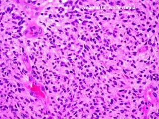
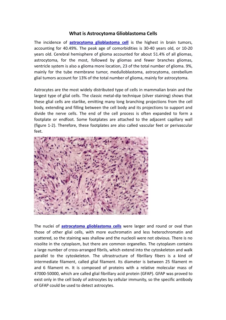
What is Astrocytoma Glioblastoma Cells The incidence of astrocytoma glioblastoma cell is the highest in brain tumors, accounting for 40.49%. The peak age of comorbidities is 30-40 years old, or 10-20 years old. Cerebral hemisphere of glioma accounted for about 51.4% of all gliomas, astrocytoma, for the most, followed by gliomas and fewer branches gliomas, ventricle system is also a glioma more location, 23 of the total number of glioma. 9%, mainly for the tube membrane tumor, medulloblastoma, astrocytoma, cerebellum glial tumors account for 13% of the total number of glioma, mainly for astrocytoma. Astrocytes are the most widely distributed type of cells in mammalian brain and the largest type of glial cells. The classic metal-dip technique (silver staining) shows that these glial cells are starlike, emitting many long branching projections from the cell body, extending and filling between the cell body and its projections to support and divide the nerve cells. The end of the cell process is often expanded to form a footplate or endfoot. Some footplates are attached to the adjacent capillary wall (figure 1-2). Therefore, these footplates are also called vascular feet or perivascular feet. The nuclei of astrocytoma glioblastoma cells were larger and round or oval than those of other glial cells, with more euchromatin and less heterochromatin and scattered, so the staining was shallow and the nucleoli were not obvious. There is no nisolite in the cytoplasm, but there are common organelles. The cytoplasm contains a large number of cross-arranged fibrils, which extend into the cytoskeleton and walk parallel to the cytoskeleton. The ultrastructure of fibrillary fibers is a kind of intermediate filament, called glial filament. Its diameter is between 25 filament m and 6 filament m. It is composed of proteins with a relative molecular mass of 47000-50000, which are called glial fibrillary acid protein (GFAP). GFAP was proved to exist only in the cell body of astrocytes by cellular immunity, so the specific antibody of GFAP could be used to detect astrocytes.
Fibrous astrocytes (fibrous astrocyte) distribution in the cerebral cortex of the spinal cord, protuberant, slender and branch less, cytoplasm contains a large number of glial wire, also known as a spider cells (spiders cell); Protoplasmic astrocytes (protoplasmic astrocytes), mostly distributed in gray matter, have short, thick, branching processes. There are few glial filaments in cytoplasm, also known as mossycell.Under electron microscope, the nucleus of astrocytes was absent, the cytoplasm was clear, the free ribosomal protein body and rough endoplasmic reticulum were few, glycogen particles were abundant, and there were a large number of glial filaments.Fibroid astrocytes have long cylindrical protuberances, while protoplasmic astrocytes have thin lamellar protuberances that often enclose nerve cells and their synapses (not extending into the synaptic space).There is a layer of substrate between the foot plate of astrocytes and the vascular endothelial cells. There are gap junctions between adjacent astrocytoma glioblastoma cells and between adjacent footplates. The intercellular space between astrocytes is narrow, only about 3nm, and contains tissue fluid.Gap joints, also known as joint pipe joints or joint membranes (nexus), consist of a large number of connexons arranged regularly in a flat plane. Each connosome is in turn composed of six subunit Mosaic proteins called connexins or connexins. A central tubule (centralcanaliculum) connects adjacent cells to the center of the small body. The gap junctions between astrocytes are mainly composed of zygosin 43(CX43). The gap junction of oligodendrocytes was formed by CX32.