Download
1 / 86
870 likes | 1.39k Vues
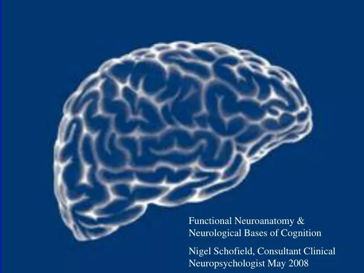
Functional Neuroanatomy & Neurological Bases of Cognition Nigel Schofield, Consultant Clinical Neuropsychologist May 2008. Evolutionary Development.

E N D
Functional Neuroanatomy & Neurological Bases of Cognition Nigel Schofield, Consultant Clinical Neuropsychologist May 2008
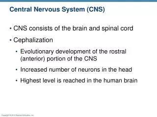
Evolutionary Development • If you examine the brain in an evolutionary perspective, this can help to understand the interlinks between form and function. The brain can be divided anatomically and functionally into three basic components.
Reptilian Brain • Corresponds to the brainstem • Consists of the medulla, pons, midbrain and basal ganglia • Not only responsible for vegetative functions but also for many volitional behaviours directed towards individual preservation and propagation such as feeding, drinking and sexual aggression.
Paleomammalian Brain • The primitive cortex of the limbic lobe • Subserves primitive (but distinctly mammalian behaviours) such as hoarding and parental care of offspring.
Neomammalian Brain • The neocortex • Subserves higher cognitive functioning and speech which facilitate social behaviour.
Neomammalian Paleomammalian Reptilian
Lurias Work • Simple anatomical localisation of function does not explain cognitive and behavioural complexity. • Postulated three functional units
Luria’s 3 Functional Units • Motor unit – regulates motor tone • Sensory unit – receives, processes and stores sensory information • The unit for programming, regulating and verifying action
How do the units work? • Progression from sensation through to symbolic function in each unit • Primary, secondary and tertiary areas
e.g. Damage to the Visual Parts of the Sensory Unit • Primary areas – losses of parts of the visual field • Secondary areas – may lead to poor judgement of motion, poor distance judgement, impaired colour perception • Tertiary areas – could lead to visual object agnosia
e.g. Damage in the Speech Pathways of the Motor Unit • Primary areas – dysarthria • Secondary areas – dysphasia • Tertiary areas – poor speech spontaneity
Weaknesses of Luria’s Model Does not fully explain the integration of focal brain functions because:- • Divisions between sensory and motor neurons sometimes not clear • Some functions exist several in anatomically distinct areas of the brain • There are multiple parallel functions in the brain, not simple stepwise processes
Arousal, Attention, Activation Arousal, Attention, Activation
Major Areas of Cognition • Attention and concentration • Perception • Memory • Language • Control of motor behaviour • Executive function
Attention and Concentration Three attentional networks:- • Alerting – achieving and maintaining an alert state in preparation for incoming stimuli • Orienting – selectively focusing on one or more items out of many candidate ones • Executive control – monitoring and resolving conflicts in planning, error detection and overcoming habitual actions • All dependent on the brain being “aroused”
Localisation of Attentional Networks • Alerting – frontal and parietal cortical regions particularly of the right hemisphere • Orienting – parts of the superior and inferior parietal lobe, frontal eye fields and subcortical areas such as superior colliculus of the midbrain, and pulvinar and reticulate nucleus of the thalamus • Executive control – includes midline frontal areas(especially anterior cingulate and lateral prefrontal cortex) and the basal ganglia
3 Compartments of Attention Top-down modulation from prefrontal, parietal & limbic cortices Modality & domain specific attentional modulations ( sounds, tactile stimuli, colours, motion, words, spatial targets, faces, objects, memories etc.) Bottom-up modulation from ascending reticular activating system
Components of the ARAS • Reticulothalamic cortical pathway – promotes and maintains cortical arousal by facilitating transthalamic passage of sensory material towards the cortex. • Transmitter specific pathways originating in the brainstem or basal forebrain, and projecting to the cerebral cortex – include dopaminergic projections from the raphe nucleus & noradrenergic projections from the locus coeruleus of the brainstem, and cholinergic & gabaminergic pathways originating in the nucelus basalis.
Limbic elements of attention • Anterior cingulate cortex plays a core pivotal role in attention – bilateral damage gives rise to akinetic mutism • Intralaminar nuclei of the thalamus receive inputs from the brainstem nuclei and relay info widely to the cortex, with a reciprocal feedback loop from the cortex modulating these ascending pathways via the thalamus
Cortical elements of attention • Parietal cortex involved in sustained and selective attention • Dorsolateral prefrontal cortex has a key role in divided attention
Perception How we take energy from the environment & convert it into a representation that the mind can use
Perceptual Problems • Visual field cuts • Cortical blindness • Achromatopsia – inability to discriminate between colours (medial occipito-temporal) • Hemianaesthesia • Hemineglect – ? an attentional problem • Hemispatial neglect • Hemiakinesia • Agnosias • Loss of taste and/ or smell
Types of Agnosia • Visual agnosias – inability to recognise familiar objects e.g. • Prosopagnosia – inability to recognise faces • Agnostic alexia – inability to read • Colour agnosia – inability to retrieve colour information e.g. what colour are bananas • Object agnosia – inability to name objects • Simultiagnosia – inability to recognise a whole image although individual details are recognised
Auditory agnosia – an inability to recognise auditory stimuli - Auditory/verbal information agnosia – an inability to hear words • Auditory agnosia – inability to hear environmental sounds e.g. car starting or dog barking • Receptive amusia – inability to hear music • Somatosensory agnosia (Astereognosis or tactile agnosia) - Difficulty perceiving objects by touch
Object recog, understanding & naming Visual analysis Functions Object Consequence of impairment Discrimination of Apperceptual agnosia shape, colour, location Perceptual classification Associative agnosia (modality specific) Knowledge of objects Associative agnosia (non-modality specific) Store of names Anomia & paraphasias Spoken name Object recog. Semantic system Lexicon
Apperceptive vs Associative Agnosia Basic High-level Naming Semantic visual perceptual & knowledge proc analysis identific. Apperc. / x x / Assoc. / / x x /
Prosopagnosia Face Voice, gait etc. Spoken name Visual analysis Expression, lip reading, Feature matching Face-recognition units **** Semantic system Knowledge of person Lexicon of names
Topographical Disorientation • Egocentric disorientation – an inability to represent the location of objects relative to self (often seen in conjunction with features of Balints syndrome – due to bilateral posterior parietal damage • Landmark agnosia – an inability to recognise salient environmental stimuli (buildings etc) – a form of associative agnosia due to lingual gyrus (basal occipital) damage • Anterograde spatial disorientation – an inability to create “new maps” or representaions of the environment – due to damage to the right parahippocampal gyrus
Cortical Network Memory