Download
1 / 20
220 likes | 616 Vues
The Sensory System. By: Derek, John and Chris. What is the Sensory System?.

E N D
The Sensory System By: Derek, John and Chris
What is the Sensory System? • Part of the nervous system consisting of sensory receptors that receive stimuli from internal and external environment, neural pathways that conduct this information to brain and parts of brain that processes this information.
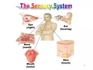
What is it made of? • Nose • Eyes • Ears • Tongue • Skin • Brain • Neurons • Receptors
The 5 Senses • Sight (vision) • Taste (gustatory) • Smell (olfactory) • Touch (tactile) • Sound (auditory)
What Triggers These Senses • Receptors • Specialized endings • Collect information about external and internal environment through a stimulus. • Each receptor is specific to a certain type of stimulus • An exception: a receptor can be activated by a nonspecific stimulus if its intensity is sufficiently high
Types of Receptors • Mechanoreceptors- respond to mechanical stimulus • Thermoreceptors- changes of temperature can stimulate these receptors • Nociceptors- brings information concerning pain • Electromagnetic receptors- rods and cones of the eye which are stimulated by changes of intensity and wavelengths of the light • Chemoreceptors- bring information concerning chemical changes in taste and smell
Receptor Potential Gating of ion channels in specialized receptor membranes allows a change in ion fluxes across the membrane, generating a graded receptor potential. The graded potential initiates an action potential, frequency and NOT magnitude of which is determined by magnitude of the graded potential. Magnitude of the receptor potential is determined by stimulus strength, summation of receptor potentials, and receptor sensitivity. The decrease in sensitivity with a constant stimulus is called adaptation.
Neural Pathways in Sensory Systems • A single afferent neuron with all its receptor endings makes a sensory unit. When stimulated, this is the portion of body that leads to activity in a particular afferent neuron is called the receptive field of that neuron. • Afferent neurons enter the CNS, diverge and synapse upon many interneurons. These afferent neurons are called sensory or ascending pathways and specific ascending pathways if they carry information about a single type of stimulus. The ascending pathways reach the cerebral cortex on the side opposite to where their sensory receptors are located. • specific ascending pathways that transmit information from somatic receptors and taste buds go to somatosensory cortex (parietal lobe), the ones from eyes go to visual cortex (occipital lobe), and the ones from ears go to auditory cortex (temporal lobe). • olfaction is NOT represented in cerebral cortex. Nonspecific ascending pathways consist of polymodal neurons and are activated by sensory units of several types. These pathways are important in alertness and arousal. • Cortical association areas, lying outside primary cortical sensory areas, participate in more complex analysis of incoming information such as comparison, memory, language, motivation, emotion etc.
Somatic Sensation Sensations from skin, muscles, bone are initiated by somatic receptors. Receptors for visceral sensations are similar.
Touch • PressureMechanoreceptors in the skin are of 2 types, rapid and slow adapting ones. • Posture and MovementMuscle-spindle stretch receptors, occurring in skeletal muscles respond to the absolute magnitude and the rate of muscle stretch. Mechanoreceptors in joints, tendons, ligaments and skin also participate. • TemperatureThermoreceptors are of two types, one that responds to an increase and the other that responds to a decrease in temperature.
Touch-Pain • Nociceptors respond to intense mechanical deformation, excessive heat etc. which cause tissue damage and many chemicals that are released by damaged cells or cells of immune system. If the initial stimulus of pain leads to an increased sensitivity to subsequent painful stimuli it is called hyperalgesia. If descending pathways inhibit the transmission of pain stimuli, it leads to a suppression of pain and this is called stimulation-produced analgesia. • If both visceral and somatic afferent converge on the same interneuron, excitation of one can lead to excitation of the other, leading to the pain being felt at a site different from the actual injured part. This is called referred pain. • Stimulating non-pain afferent fibers can inhibit neurons in the pain pathway and this therapy is called transcutaneous electric nerve stimulation (TENS). Rubbing on a painful area and acupuncture work for the same reason.
Sound • Sound Transmission in the EarOuter Ear (Pinna/Auricle) - Directs and amplifies sound waves. • External Auditory Canal –the ear canal leading from the outside to the middle ear cavityTympanic membrane (Eardrum) - Vibrates at the frequency of sound waves.Middle Ear Cavity - Filled with air. Has a movable chain of 3 bones, the malleus, incus and stapes that couple and amplifies the vibrations in the tympanic membrane to the Oval window- a membrane covered opening separating the middle ear and the Inner Ear (cochlea)Scala vestibuli - Filled with fluid Cochlear duct - Lined by the basilar membrane upon which sits the organ of Corti containing the receptor cells.
Sound cont. • Organ of CortiReceptor cells of the organ of Corti, the hair cells, are mechanoreceptors that have hairlike stereocilia. Vibration of the basilar membrane, with which the hair cells are attached, stimulates the hair cells and the pressure waves are transformed into receptor potentials. • Neural PathwaysAfferent neurons from the hair cells form the cochlear nerve. • HearingEntire audible range extends from 20 to 20,000 Hz.
Vestibular System • A series of fluid filled tubes in the inner ear that connect with each other and the cochlear duct containing hair cells that detect changes in motion 2 Parts: • Semicircular CanalsDetect angular acceleration during rotation of the head along the three axes. • Utricle and SacculeProvide information about linear acceleration and changes in head position relative to gravity. • Vestibular InformationInformation from hair cells in the vestibular apparatus is transmitted to the parietal lobe and is integrated with information from other parts of body, leading to sense of posture and movement. Unexpected inputs from the vestibular system leads to vertigo or motion sickness.
Taste • Humans perceive five types of taste: bitter, sour, salty, sweet, and umami. • Taste enables vertebrates to distinguish nutritious from noxious substances • Neurons from the tongue travel to the medulla oblongata and to the thalamus. From here, they are routed to the appropriate areas of the cerebral cortex.
Taste cont. • Taste buds are found primarily in the tongue papillae. The tongue contains 4 types of papillae, the most common type, filiform, are thin and wire shaped and do not contain taste buds. On the dorsal, anterior border of the tongue are mushroom shaped papillae, fungiform, these have taste buds located near the middle or in a cleft of the papillae. The foliate papillae are leaf shaped with taste buds on the side of the papillae, and these are along the border. The circumvallate papillae contain taste buds along the sides of whorls and are located in the posterior third of the tongue in the shape of a V. Taste buds are also located in the oral mucosa of the palate and epiglottis.
Taste cont. • The taste cells are modified epithelial cells that function as sensory receptors. About 50-60 taste cells are located in pear-shaped taste buds and the taste cells through microvilli project into a taste pore. There are non-receptor basal cells which are located on the basement membrane which do not project into the taste pore. These basal cells differentiate through a series of morphological steps into a mature taste cell. The taste cells are replaced about every 10 days. • In the past, it was believed that taste buds for sweet only existed on the tip of the tongue and for bitter were in the posterior region, but currently it is known that the tongue does not have regional differences for the taste qualities. Furthermore, taste cells can respond to the stimuli of many different taste molecules that are sweet, salt, sour or bitter
Smell • The sense of smell, called olfaction, involves the detection and perception of chemicals floating in the air. Chemical molecules enter the nose and dissolve in mucous within a membrane called the olfactory epithelium. In humans, the olfactory epithelium is located about 7 cm up and into the nose from the nostrils. • Hair cells are the receptors in the olfactory epithelium that respond to particular chemicals. These cells have small hairs called cilia on one side and an axon on the other side. In humans, there are about 40 million olfactory receptors; in the German Shepherd dog, there are about 2 billion olfactory receptors. • The electrical activity produced in these hair cells is transmitted to the olfactory bulb. The information is then passed on to mitral cells in the olfactory bulb. • The olfactory tract transmits the signals to the brain to areas such as the olfactory cortex, hippocampus, amygdala, and hypothalamus. Many of these brain areas are part of the limbic system. The limbic system is involved with emotional behavior and memory. That's why when you smell something, it often brings back memories associated with the object.
Sight • The Structure of the Eye • Aqueous Humor- Clear, watery fluid found in the anterior chamber of the eye. • Choroid- Layer of blood vessels that nourish the eye; also, because of the high melanocytes content, the choroid acts as a light-absorbing layer. • Cornea- Transparent tissue covering the front of the eye. Does not have any blood vessels; does have nerves. • Iris- Circular band of muscles that controls the size of the pupil. The pigmentation of the iris gives "color" to the eye. Blue eyes have the least amount of pigment; brown eyes have the most. • Lens- Transparent tissue that bends light passing through the eye. To focus light, the lens can change shape by bending. • Pupil- Hole in the center of the eye where light passes through. • Retina- Layer of tissue on the back portion of the eye that contains cells responsive to light (photoreceptors) • Rods- Photoreceptors responsive in low light conditions. • Cones- Photoreceptors responsive to color and in bright conditions. • Sclera- Protect coating around the posterior five-sixths of the eyeball • Vitreous Humor- Clear, jelly-like fluid found in the back portion of the eye. Maintains shape of the eye.
Sight cont. • How we see an object • Light enter eye through cornea • Light ray moves through pupil which is surrounded by iris to keep out extra light. • Light rays move through crystalline lens • Light rays move through vitreous humor • Then they fall on the retina, which processes and converts incident light to neuron signals are transmitted through the optic nerve • Neuron signals move through visual pathway- optic nerve- optic chiasm- optic tract- optic radiations- cortex • The neuron signals reach the visual cortex and its radiations for the brain’s processing • The visual cortex interprets the signals as images and along with other parts of the brain, interpret the images to extract form, meaning, memory and context of the images.