Download
1 / 71
870 likes | 1.35k Vues
Sensory System. Chapter 13 Joe Pistack MS/ED. Allows us to experience the world. We experience the world through our senses. Allows us to keep track of what is happening in our bodies. Acts as a danger signal when we come in contact with a harmful stimulus. Sensory System. Senses: Seeing

E N D
Sensory System Chapter 13 Joe Pistack MS/ED
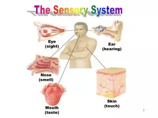
Allows us to experience the world. • We experience the world through our senses. • Allows us to keep track of what is happening in our bodies. • Acts as a danger signal when we come in contact with a harmful stimulus. Sensory System Senses: Seeing Hearing Feeling Tasting
Five types of sensory receptors: • Chemoreceptors-stimulated by changes in chemicals such as H+, calcium and food. Ex. Taste and smell. • Pain receptors or nociceptors -stimulated by tissue damage or distention. Ex. Pain. • Thermoreceptors -receptors stimulated by changes in temperature. Ex. Heat and cold Cells that detect stimuli Receptor-a specialized area of a sensory neuron that detects a specific stimulus. Ex. Receptors in the eye respond to light. Ex. Receptors in the tongue respond to chemicals in food.
Mechanoreceptors-receptors stimulated by changes in pressure or movement of body fluids. Ex. Hearing and equilibrium. • Photoreceptors-receptors stimulated by light. Ex. Sight. Receptors (continued)
Four components of a sensation: (1) Stimulus-light will stimulate sight. Absence of light, you cannot see. • (2) Receptor-light waves stimulate photoreceptors in the eye producing a nerve impulse. • (3) Sensory nerve-the nerve impulse is conducted to the occipital lobe of the brain. Sensation The conscious awareness of incoming sensory information. Ex. “Ouch”-indicates that you are aware of a painful stimulus.
Projection-the process by which the brain, after receiving a sensation, refers that sensation back to its source. Ex. Pain • Adaptation-adapting to the stimulus. Ex. When you enter a room with a strong odor, it may be overwhelming at first and then become less noticeable. Sensory receptors adapt. Sensation (continued) Two characteristics of sensation are: Projection Adaptation
Two Groups of Senses • General senses: called general or somatic. • Widely distributed throughout the body. • Include: • Pain • Touch • Pressure • Temperature • Proprioception • Special senses: • Localized within a particular organ in the head. • Include: • Taste • Smell • Sight • Hearing • Balance
Five Special Senses • Five special senses: • Smell • Taste • Sight • Hearing • balance
Pain • Pain-receptors for pain, called nociceptors consist of free nerve endings that are stimulated by tissue damage. • Pain receptors are widely distributed throughout the skin, the visceral organs, and other internal tissues.
Serves a protective function, it motivates the person to remove its cause. • Patient’s with diminished pain response are at risk . • Diabetic patients have a decreased pain sensation due to nerve damage. They are unable to feel pain in lower extremities. They require meticulous foot care. Pain
Referred Pain • Referred Pain-when pain feels as if it is coming from an area other than the site where it originates. Ex. Heart pain radiates down arm. • Analgesics-drugs to relieve pain.
Three pain triggers: • (1) Tissue injury promotes the release of certain chemicals that stimulate pain receptors. • (2) A deficiency of oxygen stimulates pain receptors. • (3) Pain may be experienced when tissues are stretched or deformed. Pain Triggers
Mechanoreceptors • Touch receptors or tactile receptors are found mostly in the skin. • They allow us to feel. • Particularly numerous in the lips, fingers, toes, tongue, and external genitalia. • Located in the skin, subcutaneous tissue, and deep tissue.
Thermoreceptors • Two types of thermoreceptors are heat and cold. • Found in free nerve endings, and specialized sensory cells beneath the skin. • Cold receptors are stimulated between 50 and 76 degrees F and heat receptors are stimulated between 76 and 112 degrees F.
Proprioception • Sense of orientation or position. • Allows you to locate your body part without looking at it. • Plays an important role in maintaining posture and coordinating body movement.
Proprioception • The receptors for proprioception are located in muscles, tendons, and joints. • Proprioceptors are also found in the inner ear, where they function in equilibrium. • The cerebellum receives sensory information from these receptors.
Sense of Smell • Olfaction-sense of smell, associated with the sensory structures located in the upper nose. • Classified as chemoreceptors, stimulated by chemicals that dissolve in the moisture of the nasal tissue.
Sense of Smell • The olfactory receptors are stimulated , the sensory impulses travel along the olfactory nerve (CN I). • The sensory information is eventually interpreted as smell within the olfactory area of the temporal lobe.
Sense of Taste • The sense of taste is also called the gustatory sense or gustation. • The taste buds are the special organs of taste. • Taste receptors are located in the tongue and are classified as chemoreceptors.
Sense of Taste • Four basic taste sensations: • Sweet-tip of the tongue. • Salty-near the tip of the tongue. • Sour-located in the middle of the tongue. • Bitter-back of the tongue.
Sense of Sight • Vision is one of the most cherished senses. • The eyes are the organs of vision. • Visual accessory organs assist with function and protecting the eye from injury.
Visual Accessory Organs • Visual accessory organs include the: • Eyebrows • Eyelids • Conjunctiva • Eyelashes • Lacrimal apparatus • Extrinsic eye muscles
Visual Accessory Organs • The eyebrows: • Patches of hair located above the eyes. • Perform a protective role. • Keep perspiration out of eyes and shade eyes from glaring sunlight. • Participate in facial expression as in the “raised eyebrow look”.
Visual Accessory Organs • Eyelids: • Also called palpebrae. • Protect the eyes. • Prevent the entrance of foreign objects. • Wash tears over the surface of the eye. • The medial inner canthus and the lateral outer canthus are the corners of the eye where the upper and lower eyelids meet.
Visual Accessory Organs • Skeletal muscle opens and closes the eyelids. • The muscle that opens the eyelid is called the levator palpebrae superioris, (levator means to raise, like an elevator). • The muscle that closes the eyelid is the obicularis oculi.
Visual Accessory Organs • Conjunctiva-thin mucous membrane that lines the inner surface of the eyelid. • The conjunctiva also folds back to cover a portion of the sclera (discussed later) on the anterior surface of the eyeball and is called the white of the eye.
Visual Accessory Organs • The conjunctiva secretes a substance that moistens the surface of the eye. • The anterior surface of the eye must be kept moist or it will ulcerate and scar. • The conjunctiva is very vascular.
Visual Accessory Organs • The “Bloodshot” appearance of the eyes is caused when the blood vessels dilate. • Eye drops that get the red out, cause the blood vessels of the conjunctiva to constrict. • Conjunctivitis-”pink eye” inflammation of the conjunctiva caused by a bacterial infection.
Visual Accessory Organs • Eyelashes: • Line the edges of the eyelid and help to trap dust. • Touching the eyelash stimulates blinking. • Sty or hordeolum -infection of the hair follicle caused by staph.
Visual Accessory Organs • Lacrimal Apparatus: • Composed of the lacrimal gland and a series of ducts called tear ducts. • The lacrimal gland is located in the upper lateral part of the orbit and secretes tears which flow across the surface toward the nose.
Visual Accessory Organs • Tears drain through small openings called lacrimal puncta and then into the lacrimal sac and nasolacrimal ducts. • The nasolacrimal ducts eventually empty into the nasal cavity.
Visual Accessory Organs • Normal tears flow to the back of the throat and are swallowed. • Increase in tears, as in crying, nose begins to run. • Tears perform several functions, moisten, lubricate and cleanse the surface of the eye. • Tears contain losozyme which helps destroy pathogens and prevent infection
The Eyeball • Has a spherical shape and is approximately ¾ to 1 inch in diameter. • Most of the eyeball sits within the bony orbital cavity of the skull, partially surrounded by a layer of orbital fat. • Composed of three layers: sclera, choroid, and the retina.
The Sclera • The outermost layer. • Made up of tough, fibrous, connective tissue that covers most of the eyeball. • Helps contain the contents of the eye. • Shapes the eye and is the site of attachment for extrinsic muscles.
The Cornea • Cornea-transparent extension of the sclera. • Covers the area over the iris. • Cornea is avascular and transparent, light rays can go through this structure. • Called the window of the eye
The Cornea • The cornea has a rich supply of nerve fibers , sensitive to touch. • Corneal reflex-protective function, if the surface of the cornea is touched lightly, causes blinking.
The Choroid • The Choroid is the middle layer of the eye located between the retina and the sclera. • Highly vascular. • Attached to the innermost layer of the retina. • Extends toward the front of the eyeball to form the ciliary body.
The Choroid • The ciliary body secretes a fluid called the aqueous humor. • Gives rise to the ciliary muscles. • Iris-most anterior portion of the choroid, the colored portion of the eye.
The Pupil • Pupil-opening or hole in the middle of the iris. • Pupil size is regulated by two sets of intrinsic muscles located in the iris. • Iris regulates the amount of light entering the eye.
The Retina • Innermost layer of the eyeball. • Nervous layer containing visual receptors which are sensitive to light, called photoreceptors. • Two types of photoreceptors are rods and cones.
The Retina • Rods, scattered throughout but most abundant along periphery. • Cones, most abundant in the central portion of the retina. • Fovea centralis area that contains the highest concentration of cones.
The Retina • The Optic Nerve forms where the neurons of the retina converge in a small circular area in the back of the eye called the optic disc. • Because there are no photoreceptors there, images formed there are not seen by the brain. This area is called the “Blind Spot”
Cavities of the Eye • Anterior Cavity • Located between the Lens and Cornea • Filled with Aqueous Humor • Aqueous Humor helps maintain shape of anterior eye and nourishes the cornea • Posterior Cavity • Larger and located between the Lens and Retina • Filled with Vitreous Humor • Vitreous Humor pushes on the retina creating good contact with the Choroid ensuring a rich supply of oxygenated blood
Eye Muscles • Extrinsic eye muscles: • Skeletal muscles. • Located outside the eye. • Move the eyeball in various directions. • Eyes move together in a coordinated way.
Seeing • Light waves enter your eye, are refracted and focused on the photoreceptors of the retina. • Photoreceptors of the retina translate light impulses, to a nerve impulse, which is then transmitted from the retina, along the optic nerve to the occipital lobe of the brain.
Muscles of the Eye • Strabismus: • Also called cross-eyed. • Eyeballs are not aligned, do not focus on a desired point.
Muscles of the Iris • Iris contains two muscles: • Radial muscle • Circular muscle • Contraction of the radial muscle causes the pupil to dilate. • Contraction of the circular muscle causes pupil to constrict.
Mydriasis-sympathetic nerve stimulation that causes pupillary dilation. • Mydriatic agents-drugs that cause the pupils to dilate. Mydriasis