Download
1 / 38
550 likes | 2.43k Vues
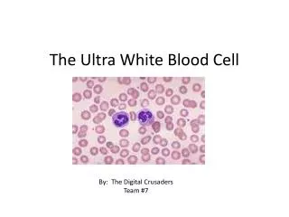
White Blood Cell Disorders. Jonathan Ben-Ezra, M.D. Professor of Pathology MCV Campus of VCU. White Blood Cells. Infection fighting cells of blood Excess causes tissue infiltration/leukostasis 4.4- 5.9 x 10 6 /L (M) 3.8 -5.2 x 10 6 /L (F) Several Types of Cells Granulocytes Lymphocytes

E N D
White Blood Cell Disorders Jonathan Ben-Ezra, M.D. Professor of Pathology MCV Campus of VCU
White Blood Cells • Infection fighting cells of blood • Excess causes tissue infiltration/leukostasis • 4.4- 5.9 x 10⁶/L (M) • 3.8 -5.2 x 10⁶/L (F) • Several Types of Cells • Granulocytes • Lymphocytes • Monocytes
Granulocytes • Produced in the Bone Marrow • Neutrophils • Life span of 8 hours • Bacteriocidal • Eosinophils • Basophils • Approximately 2/3 of WBCs in Blood
Granulocytes • Increased in Bacterial Infection • May see early forms (left shift) • Increased in Myeloid Leukemia
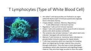
Lymphocytes • B cells • Antibody production • T cells • Fight viral infection • Morphologically can’t distinguish between the two • Approximately 30% of WBCs in PB • Increased in viral infections and lymphoid leukemia
Monocytes • Become tissue macrophages • Involved in phagocytosis • Approximately 15% of WBCs in PB
Leukemia • Neoplastic Proliferations of WBCs in Blood and Bone Marrow • Anemia, infection, bleeding • Acute Leukemias • Blast (precursor) cells • Rapidly fatal if not treated • Chronic Leukemias • More mature cells • Longer life expectancy
Acute Lymphoblastic Leukemia • Proliferation of lymphoblasts • anemia, thrombocytopenia, increased WBC • lymphadenopathy/splenomegaly • B- or T-cell • flow cytometry • TdT+ • Most common leukemia of childhood
Acute Myelogenous Leukemia • Proliferation of myeloblasts • anemia, thrombocytopenia, increased WBC • Myeloid, monocytic, RBC, or megakaryocytic • flow cytometry • myeloperoxidase +, TdT- • Auer rod • Over age of 20
Chronic Myelogenous Leukemia • 1 of myeloproliferative diseases (PV, ET) • Proliferation of more mature granulocytes • normal to increased platelet count • anemia • Splenomegaly • t(9;22) (bcr-abl) (Philadelphia chromosome)
Chronic Myelogenous Leukemia • Long chronic phase • Blast crisis • Hydroxyurea, interferons • Bone marrow transplantation
Myelofibrosis • Marrow becomes fibrotic • extramedullary hematopoiesis • dry tap • Teardrop RBC • Myeloproliferative, toxin, infection
Chronic Lymphocytic Leukemia • Proliferation of small mature B-lymphocytes • flow cytometry (monoclonal Kappa or lambda) • Lymphadenopathy • relationship to small lymphocytic lymphoma • May have Ab production and AIHA • 50% 5-year survival
Multiple Myeloma • Neoplasm of plasma cells • monoclonal protein in serum (SPEP) • Proteinuria (Bence-Jones) (UPEP) • Lytic lesions in bones • fractures • Anemia, increased globulin • Rouleaux formation • Renal failure/ amyloidosis
Infectious Mononucleosis • Acute infection with EBV • Adolescent/Young Adult • Fever, sore throat, splenomegaly, fatigue • Heterophile antibodies • Usually self limited
Lymphomas • Nodal based malignant proliferations of lymphoid cells • Hodgkin’s disease • Non-Hodgkin’s lymphoma • follicular vs. diffuse • small vs. large cell • Staging
Low grade NHL • Small lymphocytic lymphoma • nodal counterpart for CLL • Follicular lymphomas • B-cell • t(14;18) • failure of apoptosis
High Grade NHL • May be nodal or extranodal • May be T- or B-cell • Adults or pediatric • Large cell lymphoma • Burkitt’s lymphoma • t(8;14) • related to EBV • Lymphoblastic lymphoma
Extranodal Marginal Zone B-cell Lymphoma of Mucosa-Associated Lymphoid Tissue (MALT) • Proliferation of mucosal associated lymphoid tissue • Small lymphoid cells, with/out abundant cytoplasm • May transform to large cell lymphoma • Median survival of 8 years
Extranodal Marginal Zone B-cell Lymphoma of Mucosa-Associated Lymphoid Tissue (MALT)
Hodgkin Lymphoma • Bimodal age distribution • Related to EBV • Reed Sternberg cell • other background inflammatory cells • Radiation, chemotherapy, or BMT
Hodgkin Lymphoma • Nodular Lymphocyte Predominant • Nodular Sclerosis • Mixed Cellularity • Lymphocyte Depleted